What is a Ptosis?

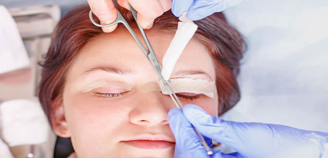
Ptosis (toh-SIS) is short for blepharoptosis. Ptosis is known as the dropping or falling of the upper eyelid over the eye. This happens when the border of the upper eyelids fall into a lower position than normal. In mild cases the upper eyelid might just droop a little, which is barely noticeable. However, in more severe cases it can cover the pupil which blocks the light from entering the eye and this can affect vision. Ptosis can present in one eye or both eyes. It can happen in both children and adults. The great news is this eye condition can be treated to improve vision as well as appearance cosmetically which helps improve the quality of life.

The three main reasons for droopy eyelids are dropping of the eyebrow, excessive skin of the upper eyelid and the dropping of the actual upper eyelid by itself. Ptosis can be congenital or acquired. Congenital ptosis normally presents at birth and occurs when the muscle that lifts the eyelid known as levator muscle does not develop well. Common signs of ptosis seen in children is a drooping eyelid or the upper eyelid creases are not aligned evenly with each other. Usually a child with ptosis will try to move their head back, lift up their chin, or raise their eyebrows in order to see well. Gradually, these will cause a child to have head and neck problems. In more severe cases, droopy lids that cover the pupil will affect the vision as well. In the long term, it can cause amblyopia or lazy eye. Some children with ptosis can develop astigmatism, strabismus or crossed eyes.

Acquired ptosis usually happens in later life in adults due to aging where the levator muscle stretches or separates away from the eyelid. This can happen by an eye injury (trauma) as well. Sometimes, acquired ptosis can also present after an eye surgery such cataract, botox injection, contact lens wear, tumours, in neurological disorder or systemic disease. Therefore, droopy eyelids can be a warning sign of severe medical problems.

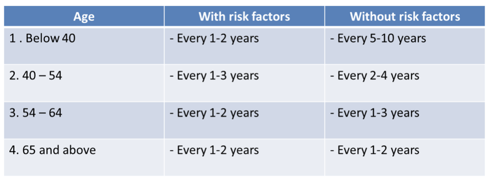
A series of eye examinations will be done by an ophthalmologist to rule out the cause of droopy eyelids for better treatment outcome. Common eye examinations include vision test and refraction especially in children to improve their vision. In moderate and severe droopy eyelids cases, surgical treatment is required for proper vision development to avoid amblyopia (lazy eyes) and poor vision. Surgical treatment for patients with normal levator muscle function is done by tightening the levator muscles that lift up eyelids and hence improve vision and appearance. In advanced cases, for patients with poor levator muscle function, the surgery is done by attaching the eyelid under the eyebrow with a sling which will help to lift the upper lid and improve vision.

Residents within Klang Valley in areas such as Cheras, Puchong, Shah Alam, Petaling Jaya, and Kepong can come visit our oculoplastic surgeon, Dr Tai Lai Yong to have their eyes examined.

Katarak

Katarak (cataract dalam bahasa Inggeris), atau juga dikenali sebagai selaput dalam adalah sejenis penyakit mata yang menyebabkan penglihatan menjadi kabur. Ianya terjadi apabila kanta semulajadi mata yang jernih bertukar menjadi keruh sehingga cahaya tidak dapat menembusnya dengan sempurna. Antara faktor utama terjadinya katarak adalah seperti proses penuaan, kecederaan mata, penyakit-penyakit seperti kencing manis (diabetes) dan penggunaan ubat steroid untuk jangka masa panjang. Katarak lebih kerap berlaku dalam kalangan orang dewasa yang berusia, namun ia juga boleh berlaku dalam kalangan kanak-kanak dan ia dikenali sebagai katarak kongenital.

Tanda & gejala

  • Penglihatan kabur tanpa rasa sakit
  • Kesukaran memandu terutamanya pada waktu malam
  • Warna kelihatan pudar atau perubahan persepsi warna
  • Kerap menukar preskripsi cermin mata
  • Silau (lampu kereta, cahaya lampu atau cahaya matahari kelihatan sangat terang)
    Penglihatan berganda pada sebelah mata (monocular diplopia)

Rawatan

Katarak boleh menganggu aktiviti harian dan menyebabkan kebutaan jika tidak dirawat. Sehingga kini tiada lagi rawatan berasaskan ubatan-ubatan yang boleh menyembuhkan katarak. Satu-satunya rawatan katarak yang berkesan adalah pembedahan. Sekiranya penglihatan cukup baik dengan menukar preskripsi cermin mata, pembedahan tidak perlu dilakukan untuk sementara waktu. Pembedahan hanya dijalankan di wad rawatan harian yang mana pesakit tidak perlu bermalam di hospital. Semasa pembedahan katarak, kanta mata yang keruh dikeluarkan dari mata dan akan digantikan dengan kanta implan intraokular (IOL) yang kekal.

Kanta intraokular (IOL)

Kanta intraokular (IOL) adalah kanta prostetik atau kanta tiruan yang digunakan untuk menggantikan kanta semula jadi yang sudah keruh semasa pembedahan katarak. Selepas implantasi kanta intraokular (IOL) ke dalam mata, ia kekal dan tidak perlu ditukar seumur hidup. Katarak tidak akan terjadi ke atas kanta intraokular (IOL). Terdapat beberapa jenis kanta yang boleh diimplan ke dalam mata semasa pembedahan. Jenis kanta yang sesuai untuk setiap orang mungkin berbeza mengikut keadaan mata yang dibedah, dan keperluan penglihatan pesakit, dan perlu dibincang bersama pakar mata masing-masing. Berikut adalah pilihan kanta IOL yang boleh digunakan untuk pembedahan katarak.[/vc_column_text][/vc_column][/vc_row][vc_row][vc_column][vc_column_text]

  1. Kanta Monofokal – menawarkan penglihatan yang baik untuk jarak jauh tetapi cermin mata diperlukan untuk membaca jarak dekat.
  2. Kanta Multifokal – menawarkan penglihatan yang baik untuk jarak jauh dan dekat.
  3. Kanta Torik – menawarkan penglihatan yang baik untuk jarak jauh dan pembetulan masalah rabun silau

Langkah-langkah yang boleh diambil untuk memperlahankan pembentukan katarak adalah:

  1. Pakai cermin mata hitam yang mempunyai perlindungan daripada cahaya UV (UV400)
  2. Amalan pemakanan yang seimbang
  3. Senaman fizikal yang berkala
  4. Elakkan merokok
  5. Elakkan penggunaan ubatan steroid tanpa pengawasan daripada doktor

Sesiapa yang menetap di Cheras, Kepong, Petaling Jaya, Puchong dan Shah Alam boleh berkunjung ke pusat kami untuk mendapat pemeriksaan mata yang menyeluruh dan nasihat daripada pakar mata kami.

Are you at risk of Glaucoma?

Glaucoma is a progressive disease that affects the optic nerve of the eye. The optic nerve is part of the central nervous system (CNS) and is the ‘cable’ that connects the eye to the brain. Once it is damaged, it cannot regenerate (repair itself). As nerve fibers of the optic nerve die slowly due to glaucoma, peripheral vision is also lost gradually. People with advanced glaucoma may experience “tunnel vision”, in which they can only see objects in the center of their visual field. The image below is an illustration of vision of a person with glaucoma.

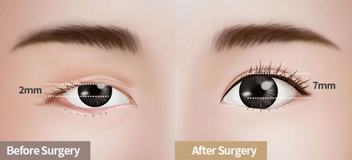
What makes glaucoma more dangerous is that the most common type of glaucoma does not show any obvious symptoms, hence goes undetected in many people. Thus, glaucoma is known as the “silent thief of sight”.

Glaucoma is one of the leading causes of irreversible blindness and therefore it is important to know the risk factors that can lead to this disease.

Strong risk factors for Glaucoma are:

  1. Advancing age (over 60 years old)
  2. Certain races are more predisposed than others (Blacks/ Hispanics/ Asians )
  3. Family history (1st degree relatives)
  4. High baseline Intraocular pressure (IOP)
  5. Suspicious optic nerve appearance / asymmetry

Possible risk factors are:

  1. Myopia (Near sightedness)
  2. Vascular disorders – Diabetes/ Hypertension/ hypercholesterolemia)
  3. Previous eye injury/ eye surgery

Other risk factors are:

  1. Long term use of steroids (eye drops/ creams/ oral/ inhalers/any other form)
  2. Migraine headaches (vasospasms)
  3. Sleep related breathing disorder (sleep apnea)
  4. Hypotension (low blood pressure)

One should have regular eye examinations as early detection is key to preventing further vision loss from glaucoma. According to guidelines by American Association of Ophthalmology (AAO), adults who have no signs or risk factors for eye disease should have a comprehensive eye examination at age 40 if they have not received one. For asymptomatic individuals without risk factors who are 40 to 54 years old, and have had a comprehensive eye examination, the recommended interval is once every 2-4 years. For those aged between 55 and 64, they should get an eye examination every 1-3 years. Anyone aged 65 or older should have an eye examination every 1-2 years, despite not having any symptoms.

By consulting a glaucoma specialist, the doctor is able to examine your eyes and determine if you have glaucoma. With the help of equipment such as the applanation tonometer, optical coherence tomography (OCT), visual field analyser, and fundus camera, the doctor is able to detect subtle changes in your optic nerve and retina.

Screening of higher-risk groups may be the most cost-effective method of reducing the volume of undiagnosed glaucoma and patient history appears to be a practical means of screening to identify the higher risk individuals.

There is a risk that if glaucoma is not detected early, irreversible loss of vision may occur, and that would affect the quality of life. Residents within Klang Valley in areas such as Cheras, Puchong, Shah Alam, and Kepong can come visit our glaucoma specialist, Datin Dr Thayanithi to have their eyes examined.

Double Eyelid Surgery

Double eyelids are visible eyelid creases. Some people were born without eyelid creases, that’s called single lid or monolid.

[vc_row][vc_column][vc_column_text]Most Asians are born with single eyelids, which is commonly due to absence of fibrous ligament connecting the tarsal to the eyelid skin. Double eyelids are the beauty standard of today and most people want double eyelids to have an appearance of larger eyes and easier to create a platform to apply make-up.[/vc_column_text][/vc_column][/vc_row][vc_row][vc_column][vc_single_image image=”4497″ img_size=”full”][/vc_column][/vc_row][vc_row][vc_column][vc_column_text]So if a person wants to have double eyelids, then can undergo a surgery that aims to create double eyelids. This procedure can be done under outpatient basis and under local anaesthesia, which means the person can go home on the same day after the procedure.[/vc_column_text][/vc_column][/vc_row][vc_row][vc_column width=”1/2″ offset=”vc_hidden-lg vc_hidden-md”][vc_single_image image=”4495″ img_size=”full”][/vc_column][vc_column width=”1/2″ offset=”vc_hidden-lg vc_hidden-md”][vc_column_text]

There are two main methods of double eyelid surgery, which are non-incisional and incisional methods. Non-incisional method is the most common method that doesn’t require removal of skin or fat. The surgeon will create a double eyelid crease by insertion of suture. The healing time for non-incisional method is faster, within 2 weeks. Furthermore, this method causes less scarring and it can be reversible. While using the incisional method, the incision will be made along the eyelid and skin or fat will be removed. People with excess skin and fat on the eyelid are suitable for the incisional method. However, this method may have a longer recovery time up to 4 weeks. There are also non-surgical techniques for creating a double eyelid appearance which is eyelid tape or glue that can be found in drugstores or beauty stores.[/vc_column_text][/vc_column][/vc_row][vc_row][vc_column offset=”vc_hidden-sm vc_hidden-xs”][vc_column_text]There are two main methods of double eyelid surgery, which are non-incisional and incisional methods. Non-incisional method is the most common method that doesn’t require removal of skin or fat. The surgeon will create a double eyelid crease by insertion of suture. The healing time for non-incisional method is faster, within 2 weeks. Furthermore, this method causes less scarring and it can be reversible. While using the incisional method, the incision will be made along the eyelid and skin or fat will be removed. People with excess skin and fat on the eyelid are suitable for the incisional method. However, this method may have a longer recovery time up to 4 weeks. There are also non-surgical techniques for creating a double eyelid appearance which is eyelid tape or glue that can be found in drugstores or beauty stores.[/vc_column_text][/vc_column][/vc_row][vc_row][vc_column offset=”vc_hidden-sm vc_hidden-xs”][vc_single_image image=”4500″ img_size=”full”][/vc_column][/vc_row][vc_row][vc_column width=”1/2″ offset=”vc_hidden-lg vc_hidden-md”][vc_single_image image=”4496″ img_size=”full”][/vc_column][vc_column width=”1/2″ offset=”vc_hidden-lg vc_hidden-md”][vc_column_text]After the surgery, there will be some discomfort and swelling of the eyelid. Medication will be prescribed after the surgery to reduce inflammation and reduce the risk of infection. For a smoother recovery, eye rubbing, eye cosmetics, strenuous or vigorous exercise should be avoided for about two weeks at least. Most individuals would be able to see a relatively natural look of the double eyelid in about three weeks after the surgery. Proper compliance of patients to the medication regime and post-operative care during recovery period will result in a better surgery outcome.

Consult with an eye doctor to find out if you are a good candidate to do the surgery. There are pros and cons of doing double eyelid surgery. During consultation you should discuss in detail about the preferred method of surgery, type of anaesthesia, expectation of the surgery, potential risk of surgery and post-operative care and recovery.[/vc_column_text][/vc_column][/vc_row][vc_row][vc_column offset=”vc_hidden-sm vc_hidden-xs”][vc_column_text]After the surgery, there will be some discomfort and swelling of the eyelid. Medication will be prescribed after the surgery to reduce inflammation and reduce the risk of infection. For a smoother recovery, eye rubbing, eye cosmetics, strenuous or vigorous exercise should be avoided for about two weeks at least. Most individuals would be able to see a relatively natural look of the double eyelid in about three weeks after the surgery. Proper compliance of patients to the medication regime and post-operative care during recovery period will result in a better surgery outcome.

Consult with an eye doctor, preferably an oculoplastic surgeon to find out if you are a good candidate to do the surgery. There are pros and cons of doing double eyelid surgery. During consultation you should discuss in detail about the preferred method of surgery, type of anaesthesia, expectation of the surgery, potential risk of surgery and post-operative care and recovery.[/vc_column_text][/vc_column][/vc_row]

Glaucoma – The Silent Thief of Sight

Does someone you know have glaucoma? It is the leading cause of irreversible blindness in the world. It affects the optic nerve, the nerve that carries visual information from your eye to your brain so that you can see. It is also a multifactorial and complex disease with specific characteristics of optic nerve damage and visual field loss.

Glaucoma and Intraocular Pressure (IOP)

Intraocular pressure is the only modifiable risk factor for this disease. Intraocular pressure (IOP) is measured with a tonometer during an eye examination. A normal intraocular pressure (IOP) is between 10-21mmHg. Although high intraocular pressure has been established as the strongest risk factor, there is no specific level of elevated eye pressure that definitely leads to glaucoma. On the contrary, there is no lower level of IOP that will absolutely eliminate a person’s risk of developing this disease. However, for specific individuals vision loss can occur when the intraocular pressure (IOP) is high (James Tsai, Glaucoma Research Foundation 2017). A person with elevated intraocular pressure is referred to as a ‘glaucoma suspect’. The term ‘glaucoma suspect’ is also used to define those patients with other risk factors such as positive family history of glaucoma or history of steroid use. Clinical risk factors include an asymmetry or cupping of the optic disc, having thin corneas with a central corneal thickness of less than 555 microns and high myopia or short-sightedness.

Early Detection

There are two types of glaucoma which are open-angle or angle-closure. Angle-closure glaucoma is more common among Asians. Patients with acute angle-closure will present to the clinic with painful red eyes, headache and periocular pain. This is also accompanied with nausea, vomiting and sudden blurring of vision. Acute-angle closure is a serious and often sudden condition that would require urgent referral to an Ophthalmologist. In open-angle glaucoma, damage to the vision tends to progress slowly over years and can ‘sneak up’ on an individual before they realize their vision is no longer normal. If you are above the age of 55, it is a good idea to see an eye doctor regularly so that it can be detected and treated early.

Watery Eyes

Watery eyes or tearing is a condition when a person produces an excess of tears and there is an overflow of tears onto the face. Tears are produced from the tear gland known as lacrimal gland and they serve few important roles to the body. They help to wash away foreign particles and dust and also act as a component of the immune system that protects us from infection. Tears also help to moisture and lubricate our eyes. An overflow of tears or too much of it on the surface of the eye can make it difficult to see.

Tears drain from the eye through the tear ducts into the nose. A blocked tear duct or a narrow duct is the most common cause of watery eyes in adults. If the tear ducts are narrowed or blocked due to inflammation or infection, the tears will not be able to drain away and thus cause excessive tearing. Some other eye conditions can also cause watery eyes. Bumps on the eyelids like stye or chalazion which are caused by infection or inflammation along your eyelids can also prompt watery eyes. When eyelashes grow inwards toward your eyes, it rubs against the cornea and causes irritation. This condition known as ingrown eyelashes or trichiasis can cause watery eyes as well.

It is also common to temporarily produce excess tears when you are in a dry environment. When you stay long in a dry environment, it prompts your tear glands to produce and release more tears which is known as reflex tearing. Exposure to pollen, pet dander, mites and fumes can cause your eyes to turn red, itchy and watery which are known as allergies. Another cause of dryness is prolonged or over wear contact lenses which can increase tear evaporation. This will evoke your tear glands into overproduction of tears and result in watery eyes. Staring at your computer screen for a long time will also result in watery eyes. When focusing too much on a computer screen, it tends to reduce our blinking rate and lead to dryness. To avoid this, we can practice the 20-20-20 rule which means that for every 20 minutes, focus on an object 20 feet away for 20 seconds. This simple action can give your eye some rest.

Watery eye treatment depends on how severe the problem is and its cause. For blocked tear ducts, surgery can create a new channel from the tear sac to the inside of the nose which allows tears to bypass the blocked part of the duct. A visit to an Ophthalmologist can help in resolving a long-standing watery eye problem.

Dr Tai Lai Yong
Consultant Ophthalmologist
Oculoplastic, Orbital & Lacrimal Surgeon

Diabetes causes blindness

Diabetes is a metabolic disease that causes high blood sugar. The hormone insulin moves sugar from the blood into your cells to be stored or used for energy. Diabetes can happen when your body either doesn’t make enough insulin or can’t effectively use the insulin it makes. Untreated high blood sugar from diabetes can damage your eyes. People with diabetes can have an eye disease called diabetic retinopathy. This happens when high blood sugar levels damage blood vessels in the retina. These blood vessels will be blocked and leaky. Sometimes abnormal new blood vessels will grow on the retina. All of these changes can make your vision become worse.

Types of diabetic retinopathy

  1. Non-proliferative diabetic retinopathy is the early stage of the disease. The blood vessels in the retina are weakened and there may be fluid leaking into the retina which may lead to swelling of the retina.
  2. Proliferative diabetic retinopathy is the advanced stage of the disease. The blood vessels might rupture and the blood may leak into the vitreous cavity, this causes patients to have acute blurry vision.

Artificial intelligence (AI) fastest way in detecting diabetic retinopathy

Previously, diabetic retinopathy was analysed and graded manually, therefore it usually takes around an hour to identify the findings. Next, these patients diagnosed as diabetic retinopathy will go through a second round of grading to ensure that the diagnosis is accurate. Artificial intelligence(AI) programme is a new technology which can deliver diagnosis within just a couple of minutes and the accuracy for AI systems is about 90% because it eliminates human inconsistencies when analysing data.

How does artificial intelligence (AI) work?

It uses selena+ software which looks out for symptoms such as bleeding,swelling and microaneurysms, which would appear as tiny red lesions in retina photography. After uploading the retinal image, the software will compare it against the dataset it has been trained on, which comprises nearly 500,000 retinal images of patients with the diseases.

OasisEye Specialists is the first Eye Centre to initiate the use of Artificial Intelligence (AI) for diabetic eye screening in Malaysia. OasisEye is ahead in providing premier eye care to its patients and collecting data to further strengthen the current AI algorithms or develop new ones.

How to prevent blindness

Diabetic retinopathy is asymptomatic at an early stage. Therefore, patients with diabetic are advised to have an annual eye screening. Early detection and treatment can prevent vision loss.

UV Light & its effects to the eye

[vc_row][vc_column][vc_column_text]The human eye is constantly being exposed to sunlight and artificial light. Natural sunlight or artificial UV rays that produce UV radiation can seriously harm the eyes, affect vision and compromise overall eye health. There are several eye diseases that are caused by long term exposure to UV radiation.

[/vc_column_text][/vc_column][/vc_row][vc_row][vc_column width=”1/2″][vc_column_text]a) Cataracts
A cataract is the clouding of the eye’s natural lens. Cataract is usually caused by aging; however, it can progress rapidly with increased exposure to UV light. This condition causes decrease in vision and glare. A cataract surgery is performed to replace the eye’s natural lens with an artificial lens.[/vc_column_text][/vc_column][vc_column width=”1/2″][vc_single_image image=”4354″ img_size=”full”][/vc_column][/vc_row][vc_row][vc_column width=”1/2″][vc_column_text]b) Photokeratitis
This condition is swelling or inflammation of the cornea due to excessive UV exposure. This is usually caused by high intensity light from welding equipment. Temporary symptoms include pain, tearing, twitching of eyelids and discomfort when exposed to bright lights. This condition requires treatment with antibiotics and artificial tears to help relieve its symptoms.[/vc_column_text][/vc_column][vc_column width=”1/2″][vc_single_image image=”4353″ img_size=”full”][/vc_column][/vc_row][vc_row][vc_column width=”1/2″][vc_column_text]c) Pterygium
A wing-shaped growth on the cornea that can show up due to prolong exposure to UV light. Those who are at risk of developing this eye condition are surfers, skiers, fisherman and farmers who spend long hours under the midday sun near the rivers, oceans and mountains. If a pterygium grows progressively, it can cause symptoms such as itchiness and discomfort. A surgery will be advised to remove it to save vision.[/vc_column_text][/vc_column][vc_column width=”1/2″][vc_single_image image=”4352″ img_size=”full”][/vc_column][/vc_row][vc_row][vc_column width=”1/2″][vc_column_text]d) Skin Cancer
Skin cancer around the eyelids have also been linked to prolong UV exposure. Melanoma, the most common type of cancer involving the eyes sometimes require surgical removal of the eyeball.[/vc_column_text][/vc_column][vc_column width=”1/2″][vc_single_image image=”4351″ img_size=”full”][/vc_column][/vc_row][vc_row][vc_column][vc_column_text]It is important to note that we do need some amount of sunlight to promote our physical and mental health in many ways. Healthy exposure to sunlight can have positive effects to a person. A little natural sunlight everyday can help you sleep well. The light-sensitive cells in the eye plays a natural role in our body’s natural wake-sleep cycles. Spending time outdoors in the natural sunlight has also been shown to prevent development of myopia or short-sightedness in kids. It is important to protect your eyes from long hours of UV exposure by using proper eyewear. Regular eye examinations with an Ophthalmologist is also important so that eye disease related to UV exposure can be detected early.[/vc_column_text][/vc_column][/vc_row]

Types of Red Eye

Red eye can be a sign of a minor irritation or a serious medical condition such as an infection. Conjunctivitis or pink eye is a common red eye in Malaysia. This eye condition is very contagious, most of the time a patient gets the red eye from either a family member or a friend who has it. It can affect one eye or both eyes can be involved as well. Common symptoms of conjunctivitis are tearing, redness, discomfort and sandy sensation in your eye. This eye disease can be resolved with medication. To avoid it spreading, patients should wash their hands frequently and avoid rubbing their eyes.

Another type of red eye is allergic eye disease which is common among children. It is associated with a history of allergic rhinitis, allergy to seafood or medication, asthma and eczema. Unlike conjunctivitis, patients will develop very severe itching associated with redness and tearing. The severe itching will lead to constant eye rubbing which eventually causes thinning of cornea (keratoconus).

Next, dry eyes are also very common in Malaysia. Those who are affected are those who are frequent contact lens wearers. Long hours of contact lens wear can cause dry eyes. Normally mild dry eyes are quite tolerable but severe dry eyes can lead to redness, frequent discomfort such as sandy feeling. In some severe cases, patient vision can be affected as well.

Red eyes can also be caused by a foreign body attaching to the eye which leads to irritation. Foreign body can be attached to the eyelid or cornea causing pain when blinking, tearing and blurry vision. Labourers are at most risk of getting this type of red eye.

The most serious cause of red eyes is corneal ulcer. It is an infection of cornea that causes thinning and perforation of the cornea as the disease progresses. It is a painful red eye. Common causes can be poor handling of contact lenses. If a corneal ulcer is not treated immediately, it can lead to severe vision loss.

An ophthalmologist will be able to assess your eye and diagnose the type of red eye you have. Some red eyes are benign while some others are serious and may need serious medical attention. Red eyes accompanied with severe pain and impaired vision should be treated immediately.

Oasis EyeQ – The Official Newsletter of OasisEye Specialists (Issue 2 / June 2021: Congratulations OasisEye Specialists)

Oasis EyeQ – The Official Newsletter of OasisEye Specialists (Issue 2 / June 2021: Congratulations OasisEye Specialists)

Congratulations OasisEye Specialists!

We are extremely honoured to have been awarded Malaysia’s Specialty Clinic of The Year by Healthcare Asia Awards. We would like to extend our sincere gratitude and appreciation to our patients, staff and well-wishers for all the support and encouragement to always strive for the best.

Also, in conjunction with cataract awareness month, here are some interesting myths and facts about cataracts. Do stay up-to-date by subscribing to the Oasis EyeQ newsletter. Find out all about the latest eye-related topics, news and events.