OasisEye Specialists

EN CN

Myopic Maculopathy

What is Myopic Maculopathy?

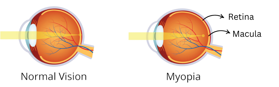
Myopic maculopathy is a condition that affects the retina and macula, which are crucial for central vision, in people with high myopia (severe nearsightedness). This eye condition occurs due to excessive elongation of the eyeball, which stretches and thins the retina, leading to degenerative changes in the macula. This condition can impair vision and, in severe cases, lead to permanent vision loss if untreated.

Comparison_of_normal_vision_and_myopia_showing_eye_anatomy._The_normal_eye_focuses_light_directly_on_the_retina,_while_the_myopic_eye_focuses_light_in_front_of_the_retina,_causing_blurred_distance_vision.

Symptoms

How can you tell if you might have myopic maculopathy? Common symptoms include

Blurry or distorted central vision

Blurry or distorted
central vision
(metamorphopsia)

Difficulty reading or recognizing faces

Difficulty reading or
recognizing faces

A dark or gray spot in central vision

Dark or blank spots
in central vision
(scotomas)

Reduced ability to see fine details

Reduced ability to
see fine details

Gradual or sudden worsening of vision

Gradual or sudden
worsening of vision

Who is at Risk?


Severe nearsightedness
(greater than -6.00 diopters)

Age-related
degeneration


Prolonged progression
of myopia


Smoking and unhealthy
lifestyle choices

What leads to Myopic Maculopathy?

High Myopia

High Myopia

Severe elongation of the eyeball stretches and thins the retina, leading to macular damage

Age

Age

Older individuals with high
myopia are more prone to
degenerative changes

Genetics

Genetics

A family history of high
myopia or macular diseases
increases the risk

Environmental Factors

Environmental Factors

Extensive near work and lack
of outdoor activities during
childhood may contribute to
high myopia

Prevention

How can you protect your eyes? Preventative measures include

Myopia Control

Myopia Control

Use myopia control in younger
individuals methods such as orthokeratology lenses, atropine
eye drops, or specialized glasses

Healthy Lifestyle

Healthy Lifestyle

● Maintain a balanced diet rich in eye-healthy nutrients (e.g., lutein, zeaxanthin, omega-3)
● Avoid prolonged screen use or near work; follow the 20-20-20 rule

Regular Eye Exams

Regular Eye Exams

Early detection of high myopia
can help prevent progression

UV Protection

UV Protection

Wear sunglasses to reduce UV-related damage to the retina
Treatment

What can be done to manage or treat myopic maculopathy?

Anti-VEGF Injections

Anti-VEGF Injections

These injections help reduce abnormal blood vessel growth and leakage
Photodynamic therapy (PDT)

Photodynamic therapy (PDT)

● PDT involves injecting a light-sensitive drug (verteporfin) and activating it with laser light to seal leaking blood vessels.

● Vitrectomy may be considered if retinal detachment or macular holes occur
Vision Aids

Vision Aids

Low vision aids and
rehabilitation in cases of
severe vision loss

Close Monitoring

Close Monitoring

Regular follow-ups with an
eye specialist to track
changes in the macula

Note: Early diagnosis and treatment can slow progression and reduce vision loss risk.

FREQUENTLY ASKED QUESTIONS

Myopic maculopathy (also known as myopic macular degeneration or pathologic myopia) is a progressive, vision-threatening eye condition affection individuals with severe high myopia (typically worse than -6.00 dioptres or an axial length greater than 26mm). It develops when progressive elongation of the eyeball stretches and thins the sclera, choroid, and retina, leading to structural damage, tissue atrophy, and abnormal blood vessel growth in the macula.

While both conditions impair central vision by affecting the macula, myopic maculopathy is caused by physical elongation of the eyeball in high myopes and often occurs in younger or middle-aged related metabolic changes, drusen accumulation, and oxidative stress in older populations (typically 50+).

Early symptoms of myopic maculopathy include:

  • Metamorphopsia: Straight lines appearing wavy, bent, or distorted.
  • Scotomas: Dark, blurry, or blank spots in central vision.
  • Decreased Contrast Sensitivity: Difficulty reading fine text or recognizing faces in dim light.
  • Sudden Vision Drop: A sudden onset of distortion or gray patches, which often indicates myopia choroidal neovascularization (CNV).

Yes, untreated myopic maculopathy can lead to permanent central vision loss. Complications such as myopic choroidal neovascularization (bleeding from abnormal blood vessels), macular hholes, or retinal detachment can destroy macular photoreceptors. However, early detection through regular dilated eye exams and prompt treatment with anti-VEGF therapy can prevent severe loss of central sight.

Retina specialists diagnose myopic maculopathy using advanced ophthalmic imaging tools, including;
1. Optical Coherence Tomography (OCT): High-resolution cross-sectional scans of retinal layers to detect fluid, macular traction, or foveoschisis.
2. Axial Length Measurement: Biometry to measure eyeball length.
3. Fundus Photography & Fluoroscein Angiography (FFA): Vissualizing retinal atrophy, lacquer cracks, and active blood vessel leakage.

Treatment depends on the specific stage and complications:

  • Anti-VEGF Injections: The gold-standard treatment for active myopic choroidal neovascularization (CNV) to stop blood vessel growth and leakage.
  • Photodynamic Therapy (PDT): Uses a light-sensitive drug and cold laser to seal abnormal leaking vessels.
  • Pars Plana Vitrectomy: Surgical treatment used if myopic traction maculopathy, macular holes, or retinal detachments occur.

No. LASIK, SMILE, and TransPRK reshape the corneal surgace to correct refractive errors, but they do not change the length of the elongated eyeball or repair the stretched retina. Patients who have undergone refractive surgery remain at the same risk for myopic maculopathy and still require routine retinal screening by an eye specialist.

Controlling myopia progression during childhood lowers the ultimate risk of pathologic high myopia. Evidence-based interventions include:

  • Low-Dose Atropine Eye Drops (0.01% to 0.05%): Slows axial elongation.
  • Orthokeratology (Ortho-K) Lenses: Overnight corneal reshaping lenses.
  • Specialized Myopia Control Glasses: Lenses with peripheral defocus technology.
  • Lifestyle Modifications: Spending at least 2 hours daily outdoors and limiting continuous near work.