Diabetic macular edema (DME) is one of the main causes of poor vision in diabetic patients. As the number of patients with diabetes mellitus is projected to increase exponentially worldwide, the incidence of DME is also expected to rise. This condition is characterized by the accumulation of fluids and thickening of the macula, the region in our eye which is most important for our central vision. The fluids are a result of leaky blood vessels damaged by the effects of diabetes. DME can occur by itself or simultaneously with diabetic retinopathy (DR), another visually-threatening eye condition from complications of diabetes mellitus.
To prevent vision loss, it is vital to manage systemic health. You are at a higher risk for DME if you have:
In its early stages, DME is often asymptomatic. Vision changes depend on the location of the edema:
At OasisEye, we use Multi-Modal Imaging to detect DME before vision fades:
| Treatment | Approach | Best For |
|---|---|---|
| Conservative Management | Careful monitoring and systemic control | Mild, non-centre-involved cases |
| Anti-VEGF Injections | Intravitreal injections (e.g., Eylea, Vabysmo) | Gold Standard for visually-threatening DME |
| Laser Therapy | Focal/Grid laser photocoagulation | Specific cases or supplementary treatment |
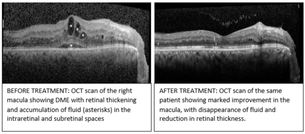
es. While DME is a serious condition, vision loss can often be reversed or stabilized with Anti-VEGF therapy and strict control of blood sugar and blood pressure. Early intervention is key to preventing permanent scarring of the macula.
No, but they are related. Diabetic Retinopathy is the general damage to retinal blood vessels, while Diabetic Macular Edema (DME) is a specific complication involving swelling in the macula. DME can occur at any stage of Diabetic Retinopathy.
Every patient is different, but many notice a reduction in blurriness within one to four weeks after their first anti-VEGF injection. However, the most significant and stable vision gains typically occur after the initial “loading phase” of three monthly injections. Consistency is key to allowing the macula to “dry” and heal.
Driving safety depends on the severity of your central vision loss. Because DME affects the macula (your “detail” vision), it can make reading road signs or seeing pedestrians difficult, even if your side vision is perfect. Your specialist will perform a visual acuity test to determine if you meet the legal vision requirements for driving in Malaysia.