Have you recently noticed sudden flashes of light or floaters in your vision? Are parts of your field of view obscured by a curtain-like shadow? These could be warning signs of retinal detachment, which is a medical emergency condition.
What are the Risk Factors of Retinal Detachment?
- Eye injury or trauma: For example, a fall, a sports accident, or a car accident.
- High myopia (short-sightedness): In high myopia individuals, the eyeball is elongated and stretched, resulting in a higher risk of getting a tear or hole in the retina.
- Diabetes: High blood sugar levels can damage the blood vessels, causing bleeding or scarring. The scar tissue can pull on the retina and cause detachment.
- Previous eye surgery: Undergoing certain types of eye surgery may increase the risk of retinal detachment, particularly if the surgery involves the vitreous or the lens of the eye.
How is Retinal Detachment Diagnosed?
- Dilated Eye Examination: The doctor will use special drops to widen your pupils, allowing them to see the retina and other structures in the back of your eye.
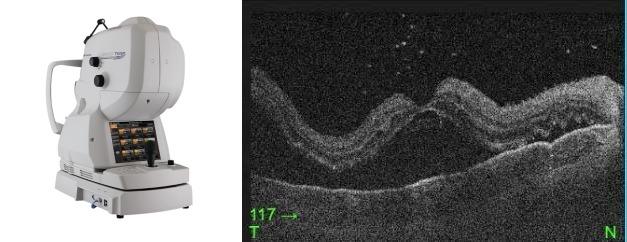
- Optical Coherence Tomography (OCT): This imaging technique provides detailed cross-sectional images of the retina, showing the separation of retina layers.
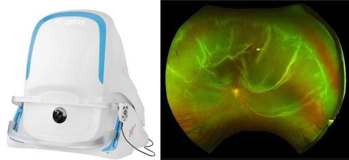
- Widefield Retinal Imaging: It provides a field of view of 200 degrees or 82% of the retina in a single capture. It helps to localise the area and the extent of the detachment.
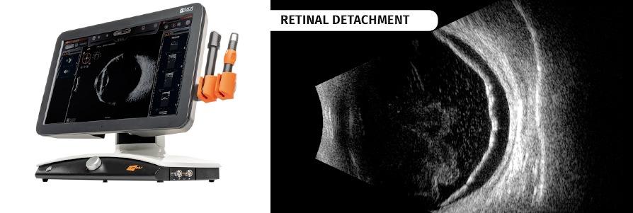
- Ultrasound: If the retina is not clearly visible (for example, due to bleeding), an ultrasound of the eye may be performed.
Treatment of Retinal Detachment:
Treating retinal detachment quickly is essential for preserving vision. The treatment approach depends on the type and extent of the detachment.
- Laser retinopexy: If a retinal tear or hole is present, a laser can be used to seal it and prevent further detachment.
- Cryopexy: It uses extreme cold to seal the tear or hole in the retina.
- Pneumatic Retinopexy: A gas bubble is injected into the eye to push the retina back into place, followed by laser or cryotherapy to seal the tear.
- Scleral Buckling: A small band (buckle) is placed around the eye to gently push the wall of the eye toward the retina, which helps to reattach it.
- Vitrectomy: This involves removing the vitreous gel and replacing it with a gas or silicone oil to help hold the retina in place.
The choice of treatment will depend on the specific circumstances of the detachment, such as its location, size, and the health of the retina.
Symptoms of Retinal Detachment:
- Floaters (dark spots or lines that seem to move with your vision)
- Flashes of light
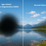
- A shadow or curtain-like effect in your vision
- A sudden loss in vision
It’s important to seek medical attention immediately if one experiences the above symptoms.
The vitreoretinal surgeons available in OasisEye Specialists include Dr Kenneth Fong, Dr Manoharan, Dr Wilson Wong who is based in Kuala Lumpur; Dr Ling Kiet Phang who is based in Johor Bahru; Dr Teh Wee Min who is based in Seremban and also Dato Dr Haslina who is based in Penang.