
Our Technologies
We are equipped with the latest ophthalmic technology to support accurate assessments and effective treatment. Our equipment is regularly updated to ensure reliability and efficiency in daily clinical practice. This enables our team to deliver safe, precise, and well-managed care, while maintaining a consistently high standard for our patients.
BIOMETRY
Biometry is the clinical process of measuring the physical dimensions of the eye to determine the correct power of an intraocular lens (IOL). It is a vital diagnostic step for patients preparing for cataract surgery or vision correction procedures.
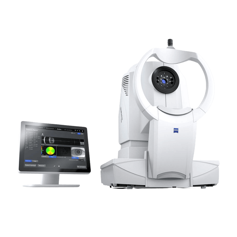
ZEISS IOLMaster
As the industry-leading flagship model, the ZEISS IOLMaster 700 utilizes advanced Swept-Source OCT technology to provide superior penetration through 99% of cataracts—including dense “white” cases—while ensuring surgical precision through a visual fixation check that confirms the patient’s focus during the scan.
Topcon Aladdin
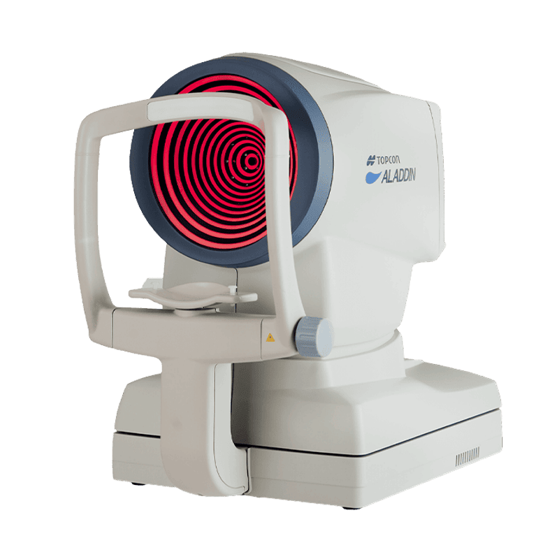
The Topcon Aladdin functions as a comprehensive, multimodal diagnostic hub that utilizes Optical Low Coherence Interferometry and integrated Placido Topography to provide 9-in-1 efficiency, offering critical corneal mapping and keratoconus screening essential for surgeons selecting Toric or premium intraocular lenses.
Topcon Aladdin
The Topcon Aladdin functions as a comprehensive, multimodal diagnostic hub that utilizes Optical Low Coherence Interferometry and integrated Placido Topography to provide 9-in-1 efficiency, offering critical corneal mapping and keratoconus screening essential for surgeons selecting Toric or premium intraocular lenses.
CORNEAL SPECULAR MICROSCOPY
Specular microscopy is a non-invasive, photographic imaging technique used to analyze the corneal endothelium, which is the single layer of cells on the innermost surface of the cornea responsible for keeping it clear and hydrated.
Topcon SP-1P
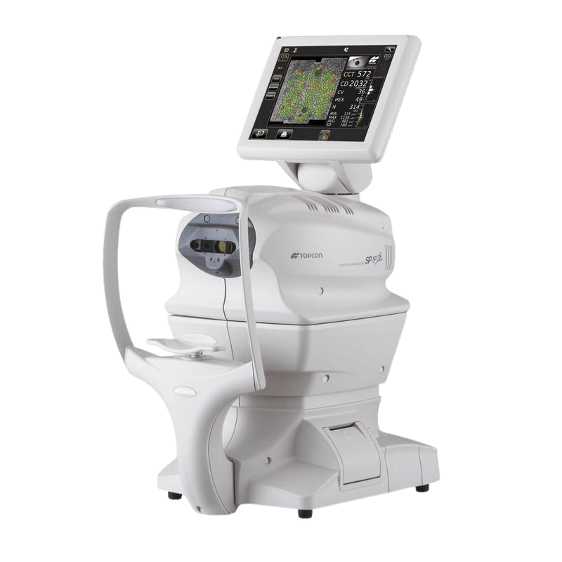
The Topcon SP-1P is a fully automated, touchscreen-driven specular microscope that utilizes a unique Panorama photography mode and integrated analysis software to provide rapid, comprehensive evaluations of endothelial cell density and corneal health, essential for pre-surgical screening and long-term corneal management.
CORNEAL TOPOGRAPHY AND TOMOGRAPHY
While corneal topography provides a 2D map of the eye’s outer surface to assess curvature and astigmatism, corneal tomography delivers a comprehensive 3D structural scan that analyzes the front surface, back surface, and total thickness to ensure safety for procedures like LASIK and Keratoconus management.
SCHWIND MS39
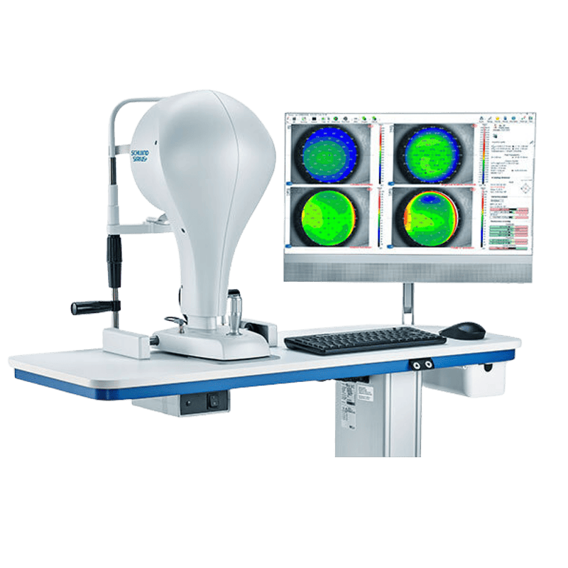
Considered one of the most advanced anterior segment workstations available, the SCHWIND MS39 combines Placido Topography with high-resolution Spectral Domain OCT to provide microscopic epithelial mapping, making it the premier tool for detecting early keratoconus and planning complex refractive repairs.
SCHWIND MS39
Considered one of the most advanced anterior segment workstations available, the SCHWIND MS39 combines Placido Topography with high-resolution Spectral Domain OCT to provide microscopic epithelial mapping, making it the premier tool for detecting early keratoconus and planning complex refractive repairs.
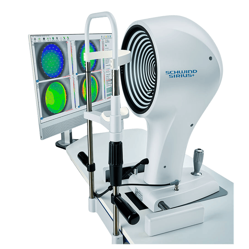
SCHWIND Sirius
As the established industry standard for clinical screening, the SCHWIND Sirius combines Placido Topography with a rotating Scheimpflug camera to deliver a fast, comprehensive 3D scan of the entire anterior segment, making it an essential tool for routine LASIK screening, cataract planning, and glaucoma diagnostics.
OCULUS Pentacam
The OCULUS Pentacam is the premier diagnostic tool for corneal tomography, using a rotating Scheimpflug camera to provide comprehensive 3D mapping of the anterior eye segment, which is vital for early keratoconus detection and ensuring the safety of refractive surgeries like LASIK.
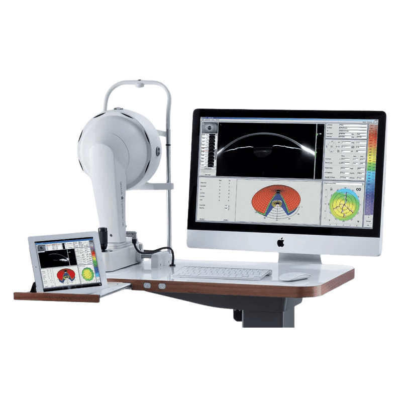
OCULUS Pentacam
The OCULUS Pentacam is the premier diagnostic tool for corneal tomography, using a rotating Scheimpflug camera to provide comprehensive 3D mapping of the anterior eye segment, which is vital for early keratoconus detection and ensuring the safety of refractive surgeries like LASIK.
DRY EYE DIAGNOSTICS AND MACHINE
Dry eye diagnostics have evolved into a comprehensive ocular surface analysis that utilizes infrared meibography, non-invasive tear film stability testing, and inflammatory biomarker detection to pinpoint the exact cause of irritation, allowing for a highly personalized treatment plan. It is also used as part of the “Digital Eye Strain Assessment Tool”.
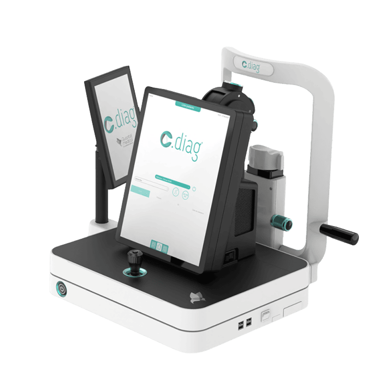
C-Diag
The Lumibird C-Diag is an integrated, non-invasive ocular surface analyzer that provides automated meibography and tear film stability metrics, enabling clinicians to objectively diagnose and monitor dry eye disease through a streamlined, slit-lamp-mounted platform.
IDRA
The IDRA is a state-of-the-art, all-in-one ocular surface analyzer that provides fully automated diagnostics—including 3D meibography and lipid layer interferometry—to deliver an objective, color-coded report for the precise management of dry eye disease.
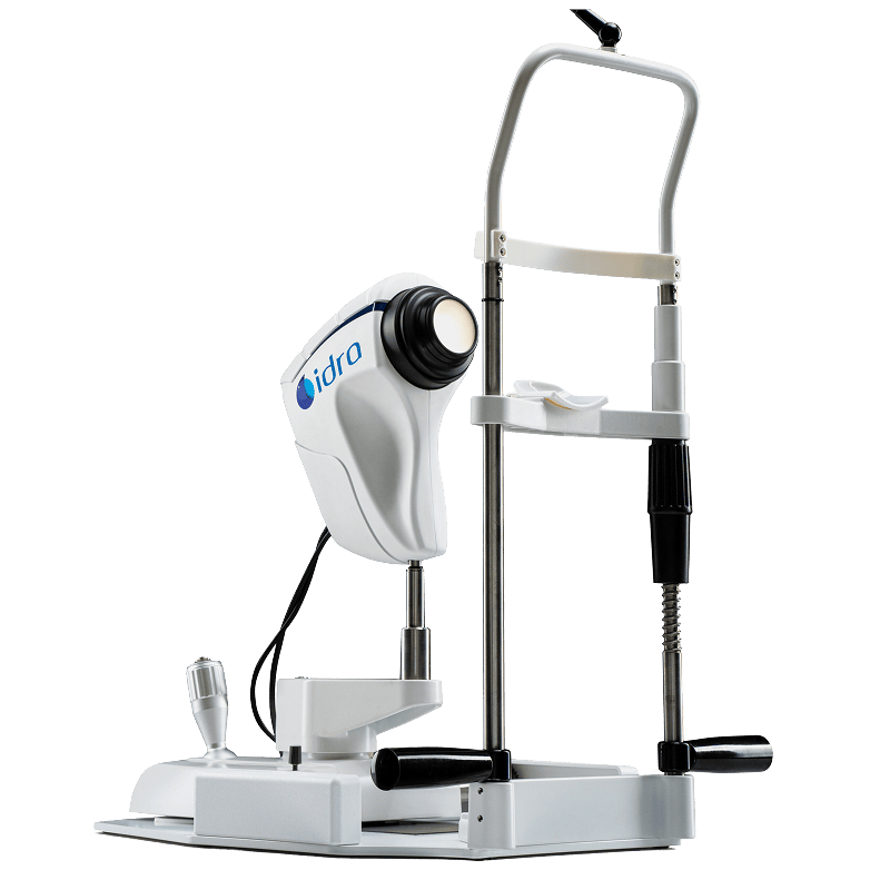
IDRA
The IDRA is a state-of-the-art, all-in-one ocular surface analyzer that provides fully automated diagnostics—including 3D meibography and lipid layer interferometry—to deliver an objective, color-coded report for the precise management of dry eye disease.
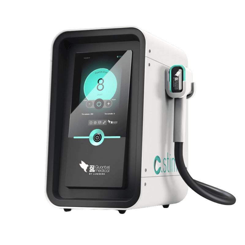
C-STIM
The C-STIM by Lumibird (Quantel Medical) is a specialized Intense Pulsed Light (IPL) system specifically engineered for the treatment of Dry Eye Disease (DED) and Meibomian Gland Dysfunction (MGD).
E-Eye
The E-Eye is a pioneering dry eye solution to provide long-lasting relief from Meibomian Gland Dysfunction by neurologically stimulating the glands to restore their natural oil production.
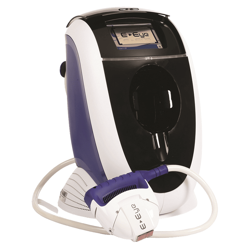
E-Eye
The E-Eye is a pioneering dry eye solution to provide long-lasting relief from Meibomian Gland Dysfunction by neurologically stimulating the glands to restore their natural oil production.
HESS CHART
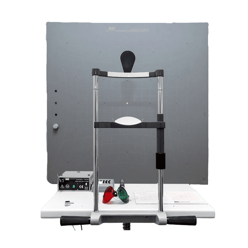
A Hess Chart (or Hess Screen Test) is a diagnostic tool used to measure and map ocular motility and the coordination of the extraocular muscles. It is essential for identifying muscle underaction (paresis) or overaction, often used in cases of strabismus or diplopia (double vision).
RETINA IMAGING
Retina imaging refers to a group of non-invasive diagnostic tests that capture high-resolution pictures of the back of the eye, including the retina, optic disc, and blood vessels. These images are essential for detecting, monitoring, and treating sight-threatening conditions like glaucoma, macular degeneration, and diabetic retinopathy.
Optos California
The Optos California is a premier ultra-widefield imaging system that captures a 200-degree view of the retina in a single scan. It’s a non-contact scan, enabling doctors to detect peripheral retinal diseases that are often missed by traditional, narrow-angle cameras. It offers multiple imaging modes, such as Color, Red-free, Fundus Autofluorescence (FAF), and Fluorescein Angiography (FA) & Indocyanine Green (ICG).
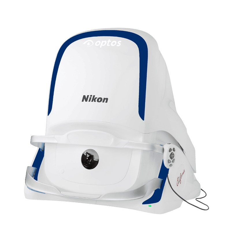
Optos California
The Optos California is a premier ultra-widefield imaging system that captures a 200-degree view of the retina in a single scan. It’s a non-contact scan, enabling doctors to detect peripheral retinal diseases that are often missed by traditional, narrow-angle cameras. It offers multiple imaging modes, such as Color, Red-free, Fundus Autofluorescence (FAF), and Fluorescein Angiography (FA) & Indocyanine Green (ICG).
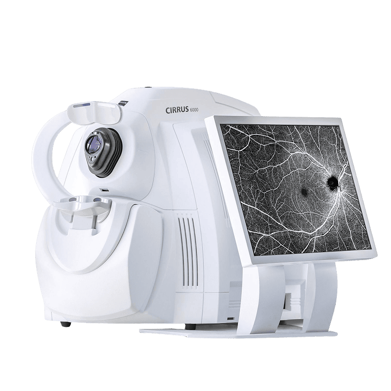
ZEISS CIRRUS™ HD-OCT
The ZEISS CIRRUS HD-OCT is an industry-leading diagnostic platform that utilizes high-speed Spectral Domain OCT and AngioPlex® technology to deliver precise 3D visualizations of the retina and optic nerve. It enables early detection and long-term monitoring of sight-threatening conditions such as glaucoma and macular degeneration. The system features “Zero-Dye Angiography” for non-invasive vascular imaging, alongside advanced AI-driven analytics, including PathFinder™, which intelligently tracks anatomical changes over time and highlights clinically relevant progression patterns—supporting more accurate and efficient decision-making.
Topcon DRI OCT Triton
The Topcon DRI OCT Triton is a premier multimodal imaging platform that leverages Swept-Source OCT technology and a 1,050nm wavelength to provide deep, high-resolution scans of the retina and choroid—deep penetration through cataracts—while combining color fundus photography and dye-free angiography in a single, efficient device.
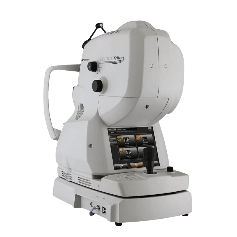
Topcon DRI OCT Triton
The Topcon DRI OCT Triton is a premier multimodal imaging platform that leverages Swept-Source OCT technology and a 1,050nm wavelength to provide deep, high-resolution scans of the retina and choroid—deep penetration through cataracts—while combining color fundus photography and dye-free angiography in a single, efficient device.
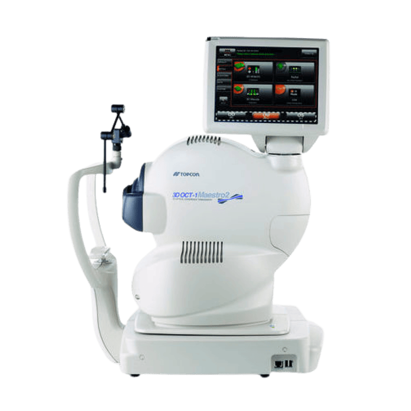
Topcon Maestro2
The Topcon Maestro2 is a user-friendly, fully automated diagnostic platform that combines Spectral Domain OCT with high-resolution true-color fundus photography to provide rapid, comprehensive 3D imaging of the retina and optic nerve at the touch of a button.
SURGICAL
Surgical equipment in ophthalmology comprises highly specialized tools—including 3D surgical microscopes, ultrasonic phacoemulsification units, and ultra-fast lasers—designed to perform micron-level maneuvers that restore vision with minimal invasiveness and rapid recovery times.
ZEISS ARTEVO 750 iOCT
The ZEISS ARTEVO 750 is a next-generation surgical microscope combining world-class optics with intraoperative OCT and AdVision® digital overlays, enabling real-time visualization of microscopic tissue layers and integrated surgical data, with OasisEye Specialists being the first eye centre in Malaysia to offer this advanced iOCT technology for enhanced precision in both cataract and retinal procedures (Vitrectomy).
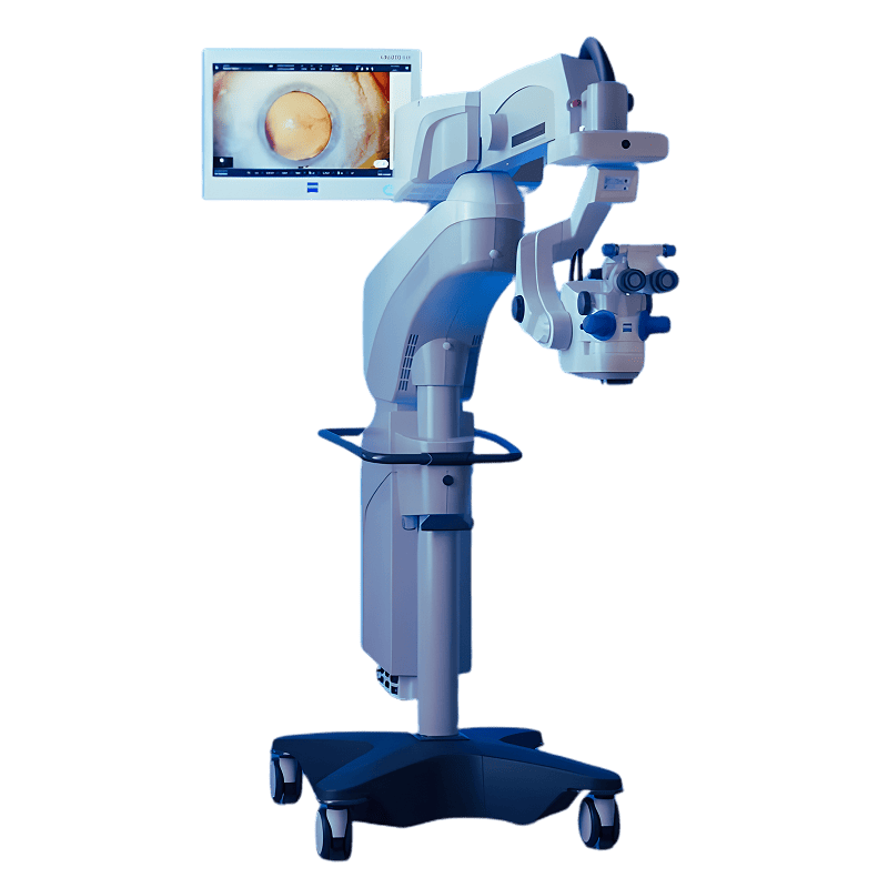
ZEISS ARTEVO 750 iOCT
The ZEISS ARTEVO 750 is a next-generation surgical microscope combining world-class optics with intraoperative OCT and AdVision® digital overlays, enabling real-time visualization of microscopic tissue layers and integrated surgical data, with OasisEye Specialists being the first eye centre in Malaysia to offer this advanced iOCT technology for enhanced precision in both cataract and retinal procedures (Vitrectomy).
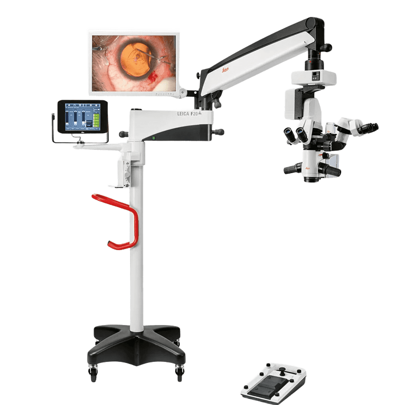
Leica Microsystems M822
The Leica M822 is a premium ophthalmic surgical microscope that pairs world-class apochromatic optics with a unique dual LED and halogen illumination system to provide surgeons with a high-definition, stable red reflex for enhanced precision in both anterior and posterior segment procedures.
Bausch + Lomb Stellaris Elite
The Bausch + Lomb Stellaris Elite® is a premium, versatile surgical platform designed for both Cataract (Anterior) and Retina (Posterior) procedures. It is particularly valued in high-volume clinics for its efficiency, advanced fluidics, and its ability to handle complex cases with minimal tissue trauma.
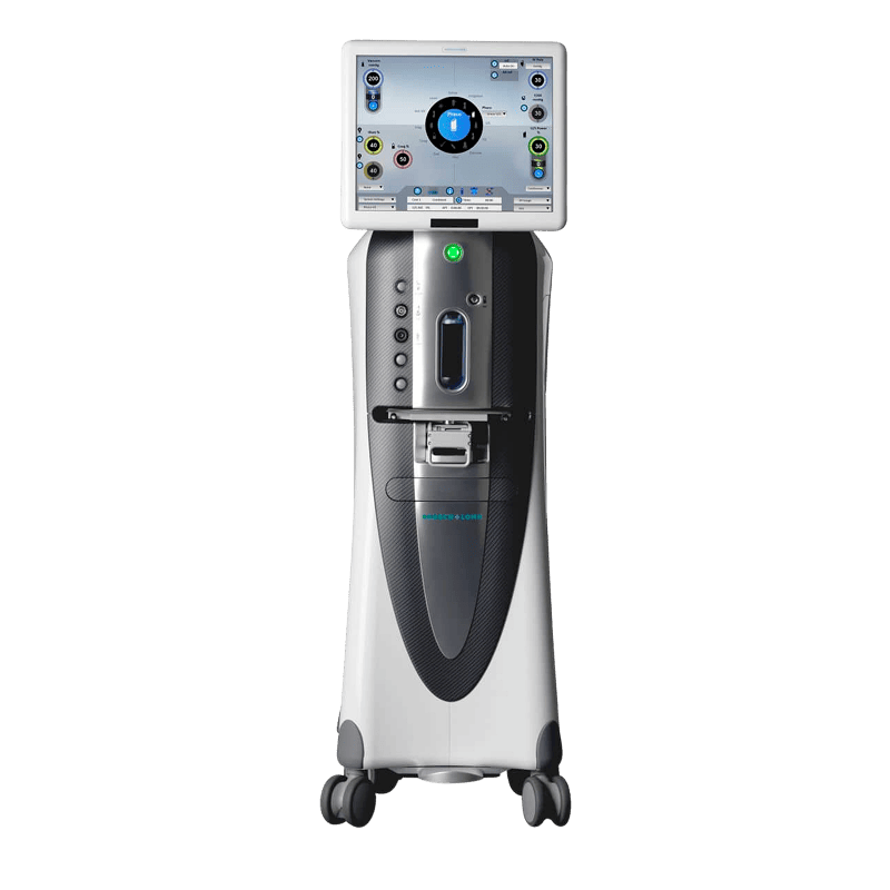
Bausch + Lomb Stellaris Elite
The Bausch + Lomb Stellaris Elite® is a premium, versatile surgical platform designed for both Cataract (Anterior) and Retina (Posterior) procedures. It is particularly valued in high-volume clinics for its efficiency, advanced fluidics, and its ability to handle complex cases with minimal tissue trauma.
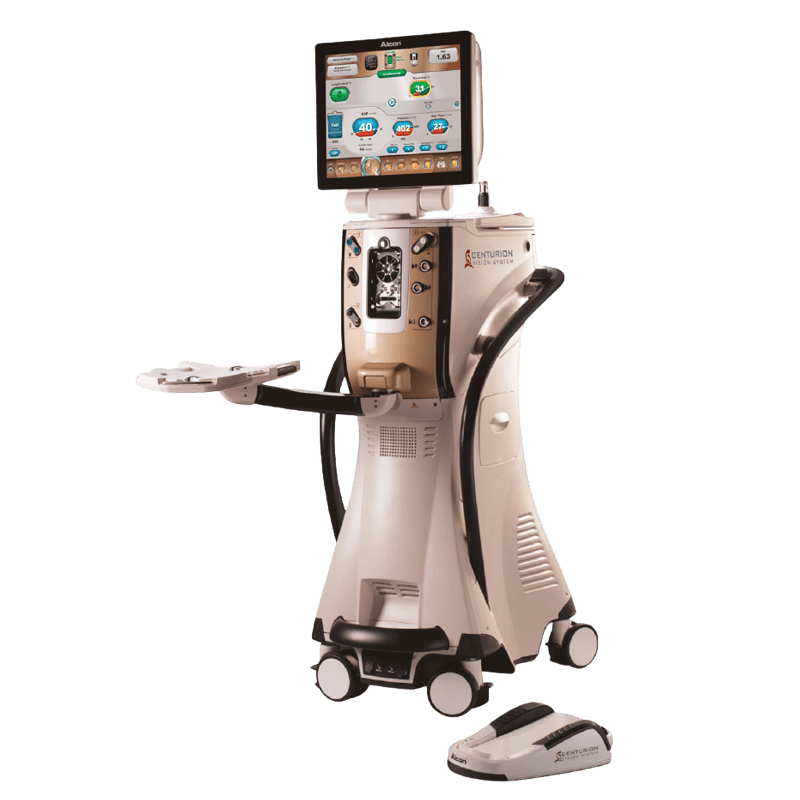
Alcon Centurion
The Alcon Centurion® Vision System is widely considered the “Gold Standard” in phacoemulsification technology for cataract surgery. It is engineered to maximize safety and efficiency by maintaining a highly stable environment inside the eye during the procedure. Often regarded as the safest cataract technology available.
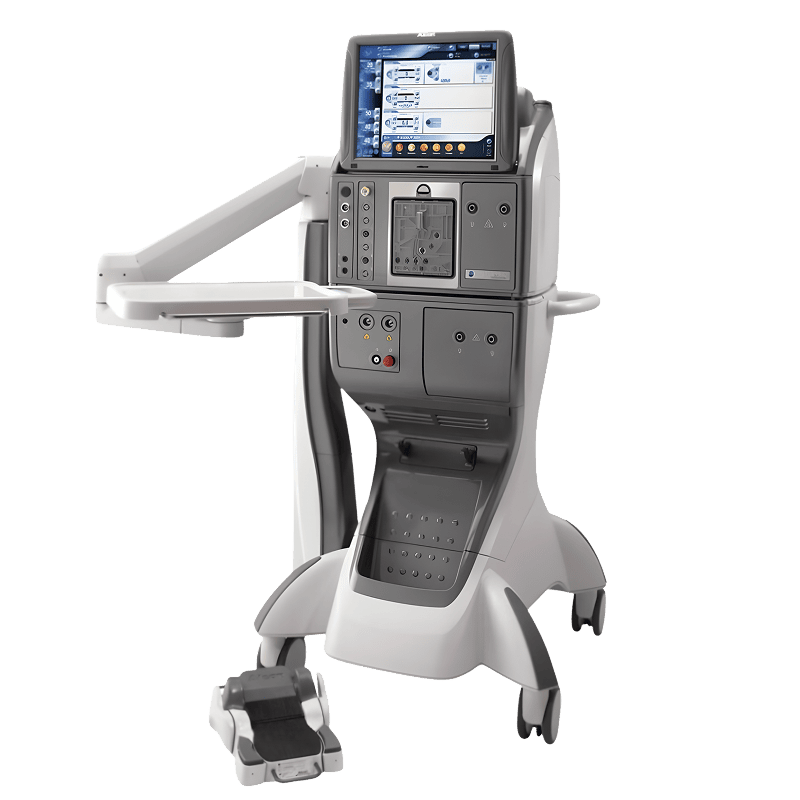
ZEISS Visumax 800
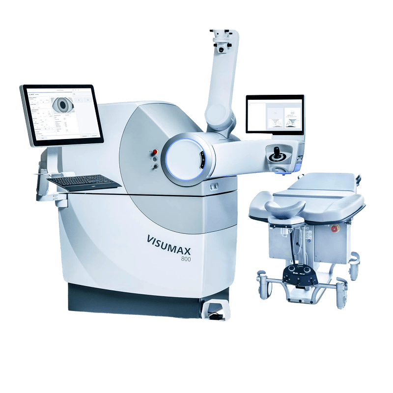
The ZEISS VISUMAX 800 is a next-generation femtosecond laser that powers SMILE® pro 2.0, utilizing a 2 MHz pulse frequency to complete laser treatments in under 10 seconds while using robotic CentraLign and OcuLign systems to deliver unprecedented precision and patient comfort. It is also a powerhouse for Femto-LASIK. In a Femto-LASIK procedure, the Visumax 800 is used for the first and most critical step: creating the corneal flap.
ZEISS MEL 90
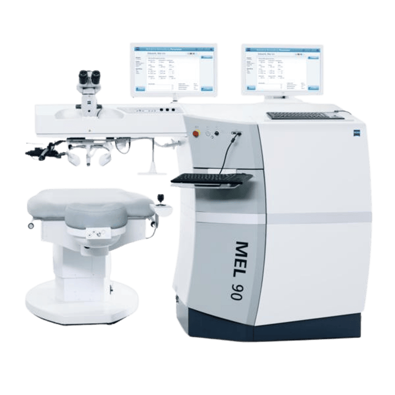
The ZEISS MEL 90 is a versatile excimer laser that utilizes the Triple-A ablation algorithm and a 500 Hz high-speed frequency to provide fast, tissue-saving, and highly predictable vision correction for LASIK and surface treatments.
PRESBYOND® 2.0 is a sophisticated physiological treatment performed with the ZEISS MEL 90 that uses “blended vision” to provide a continuous range of clear sight from near to far, making it especially suitable for individuals aged 40 and above experiencing age-related near vision changes (presbyopia), helping restore near vision and reduce dependence on reading glasses in a more natural way compared to traditional monovision.
ZEISS MEL 90
The ZEISS MEL 90 is a versatile excimer laser that utilizes the Triple-A ablation algorithm and a 500 Hz high-speed frequency to provide fast, tissue-saving, and highly predictable vision correction for LASIK and surface treatments.
PRESBYOND® 2.0 is a sophisticated physiological treatment performed with the ZEISS MEL 90 that uses “blended vision” to provide a continuous range of clear sight from near to far, making it especially suitable for individuals aged 40 and above experiencing age-related near vision changes (presbyopia), helping restore near vision and reduce dependence on reading glasses in a more natural way compared to traditional monovision.
SCHWIND AMARIS 750S
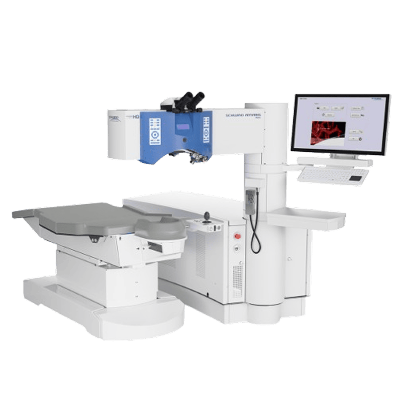
The SCHWIND AMARIS 750S is a leading “TotalTech” excimer laser that combines a 750 Hz pulse rate with active 6D eye tracking and SmartSurfACE technology to provide a completely touch-free, highly precise, and thermally optimized vision correction experience. By combining SmartPulse (for smoothness) with TransPRK (for safety) and 6D Tracking (for precision), the Amaris 750S offers a “No-Cut, No-Touch” surgery that effectively bridges the gap between the safety of PRK and the comfort of LASIK.
Bausch + Lomb TECHNOLAS TENEO 317 Model 2
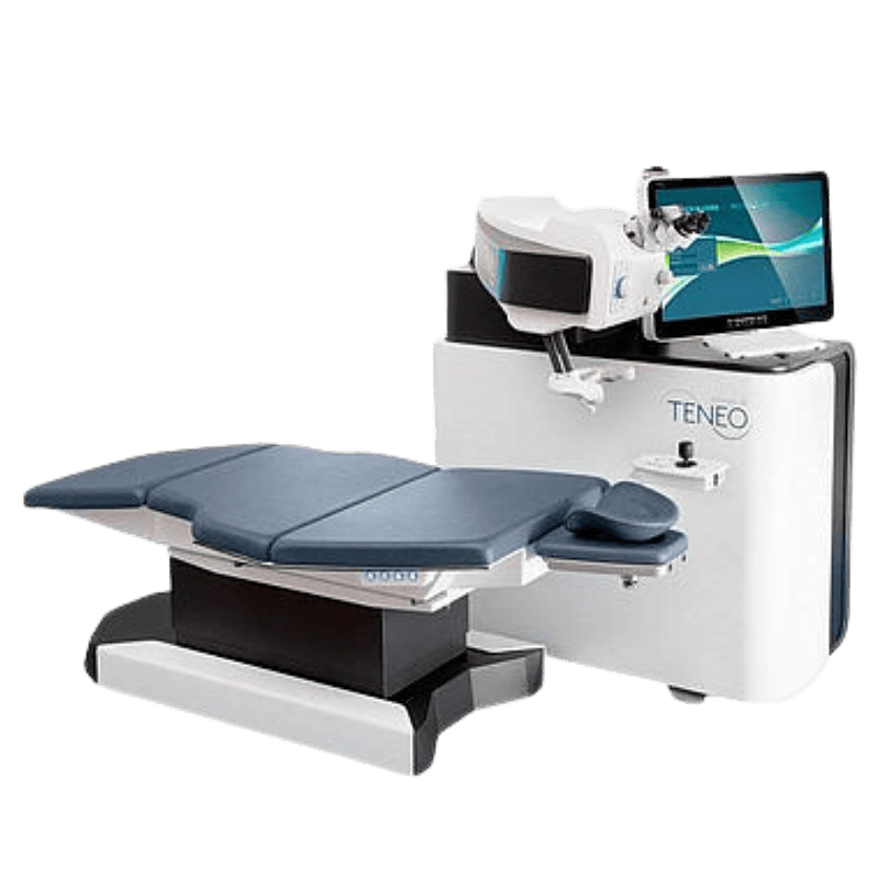
The TECHNOLAS TENEO 317 Model 2 is a fast and efficient excimer laser system that excels in customized vision correction and offers the unique SUPRACOR™ procedure, providing a non-invasive surgical solution for patients looking to correct both distance and reading vision.
Bausch + Lomb TECHNOLAS TENEO 317 Model 2
The TECHNOLAS TENEO 317 Model 2 is a fast and efficient excimer laser system that excels in customized vision correction and offers the unique SUPRACOR™ procedure, providing a non-invasive surgical solution for patients looking to correct both distance and reading vision.
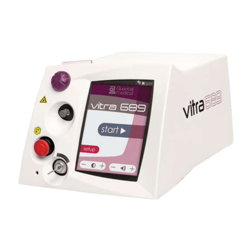
Vitra 689™ Laser
The Vitra 689 is a precision medical laser used for Photodynamic Therapy (PDT), providing a “cold” laser treatment that activates special medication to close leaky blood vessels in the eye without damaging healthy retinal tissue. Used in treatment such as Polypoidal Choroidal Vasculopathy (PCV), Central Serous Chorioretinopathy (CSCR), Choroidal Hemangioma, and pathologic myopia (with CNV).
Vitra 810™ Laser
The Vitra 810 is a multi-purpose infrared laser that specializes in non-scarring “SubLiminal” therapy, making it a powerful and gentle tool for lowering eye pressure in glaucoma and treating delicate retinal diseases.
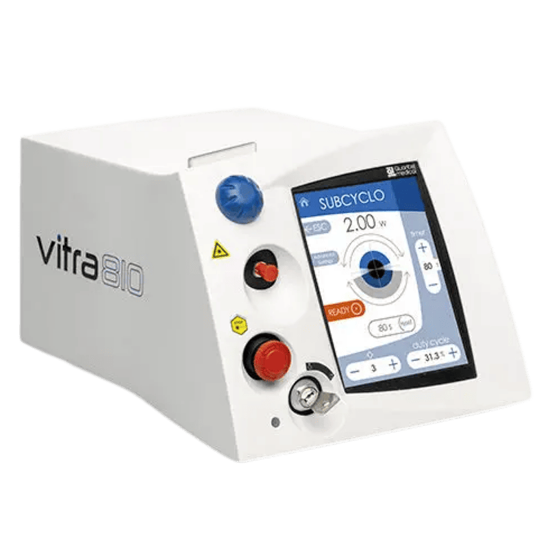
Vitra 810™ Laser
The Vitra 810 is a multi-purpose infrared laser that specializes in non-scarring “SubLiminal” therapy, making it a powerful and gentle tool for lowering eye pressure in glaucoma and treating delicate retinal diseases.
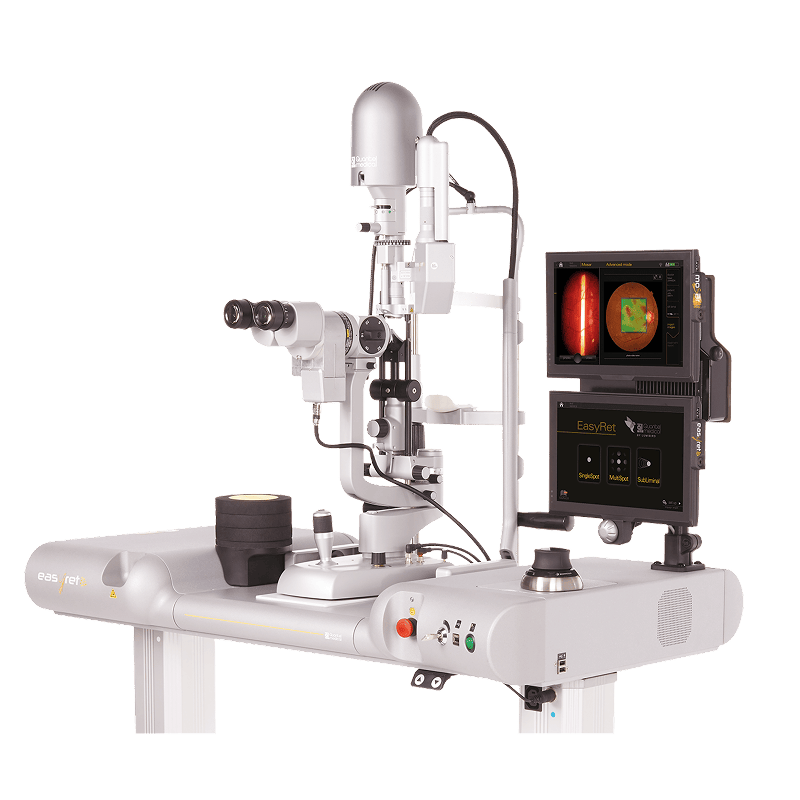
EASYRET® Yellow Laser
The EasyRet® Yellow Laser, a cutting-edge workstation designed to make retinal treatments faster, safer, and significantly more comfortable. The EasyRet’s unique 577nm yellow wavelength is much gentler on the eye; it is highly effective at treating delicate areas near the center of your vision (the macula) without damaging the surrounding healthy tissue. It is used to treat Diabetic Retinopathy, Macula Edema, Central Serous Chorioretinopathy (CSCR) and even Retina Tears.
OPTIMIS FUSION Laser
The Optimis Fusion is a versatile 2-in-1 laser system that treats the two most common causes of vision loss. It acts as a gentle pressure-reliever for glaucoma patients (SLT) and a precision “clearing” tool for patients who develop cloudiness after cataract surgery (YAG). It’s designed to be fast, painless, and exceptionally accurate.
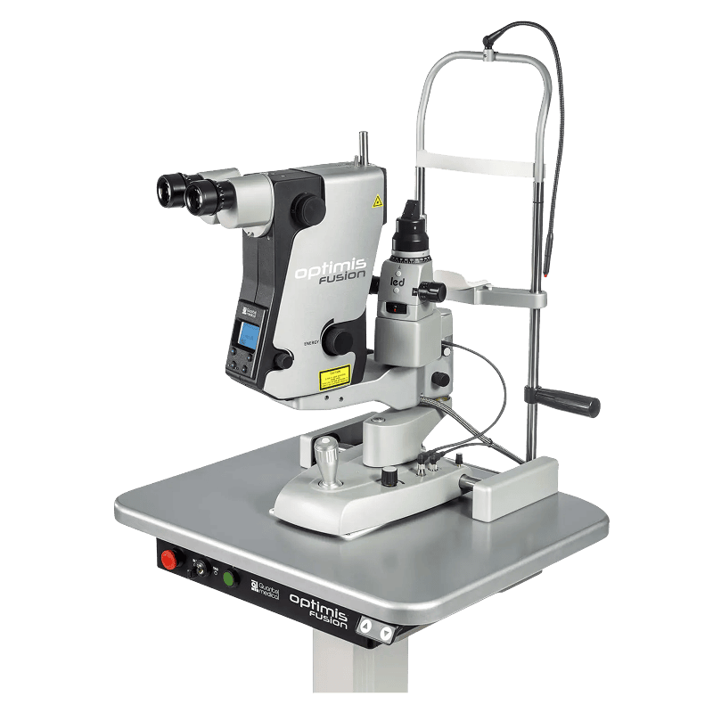
OPTIMIS FUSION Laser
The Optimis Fusion is a versatile 2-in-1 laser system that treats the two most common causes of vision loss. It acts as a gentle pressure-reliever for glaucoma patients (SLT) and a precision “clearing” tool for patients who develop cloudiness after cataract surgery (YAG). It’s designed to be fast, painless, and exceptionally accurate.
VISUAL FIELD TEST
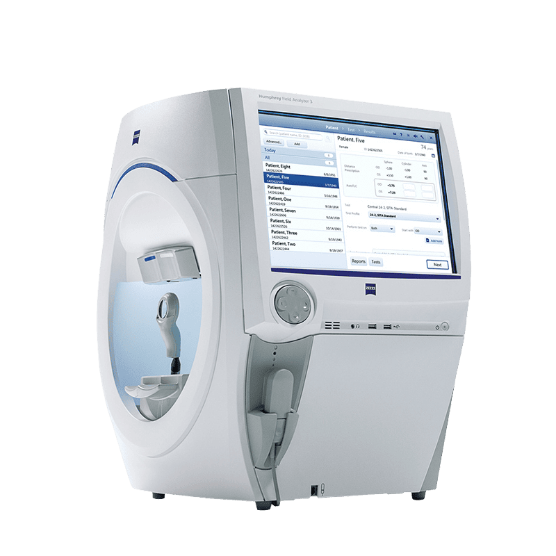
A visual field test is a subjective diagnostic exam that measures your total range of sight—including both your direct central vision and your peripheral (side) vision. It measures visual field sensitivity, mapping the entire area of vision (central and peripheral) to detect blind spots (scotoma) that the patient might not even notice yet. A vital diagnostic test for glaucoma.
ZEISS Humphrey Field Analyzer 3 (HFA)
The ZEISS Humphrey Field Analyzer 3 offers faster and more reliable visual field testing than conventional HVF systems through advanced algorithms like SITA Faster that significantly reduce test time, improved fixation monitoring for greater accuracy, seamless integration with ZEISS FORUM for precise progression tracking, and enhanced patient comfort with features such as the Liquid Trial Lens™ that eliminates the need for manual trial lenses.
Topcon Henson 9000
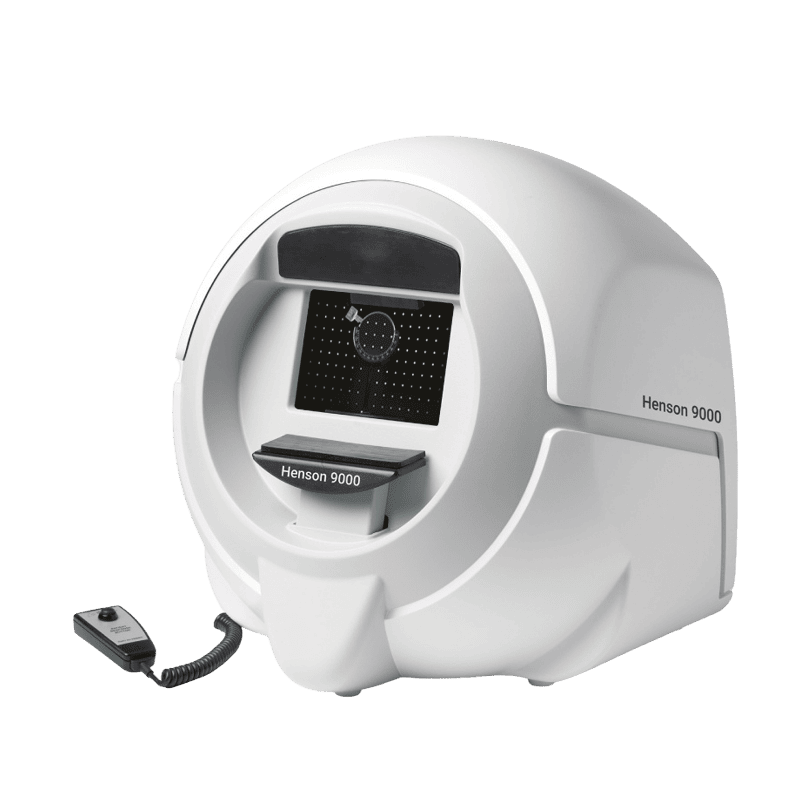
The Topcon Henson 9000 is an efficient, space-saving automated perimeter that uses the ZATA algorithm to provide both rapid glaucoma screening and detailed threshold monitoring, making it an ideal choice for clinics that prioritize workflow speed and portability.
Topcon Henson 9000
The Topcon Henson 9000 is an efficient, space-saving automated perimeter that uses the ZATA algorithm to provide both rapid glaucoma screening and detailed threshold monitoring, making it an ideal choice for clinics that prioritize workflow speed and portability.
OPHTHALMIC ULTRASONOGRAPHY
An eye ultrasound (also known as ophthalmic ultrasonography) is a non-invasive diagnostic tool that uses high-frequency sound waves to visualize the internal structures of the eye. Unlike traditional eye exams that use light (like an ophthalmoscope or OCT), an ultrasound uses sound, which allows it to “see” through obstructions like a dense cataract or blood that would otherwise block a doctor’s view.
ABSolu
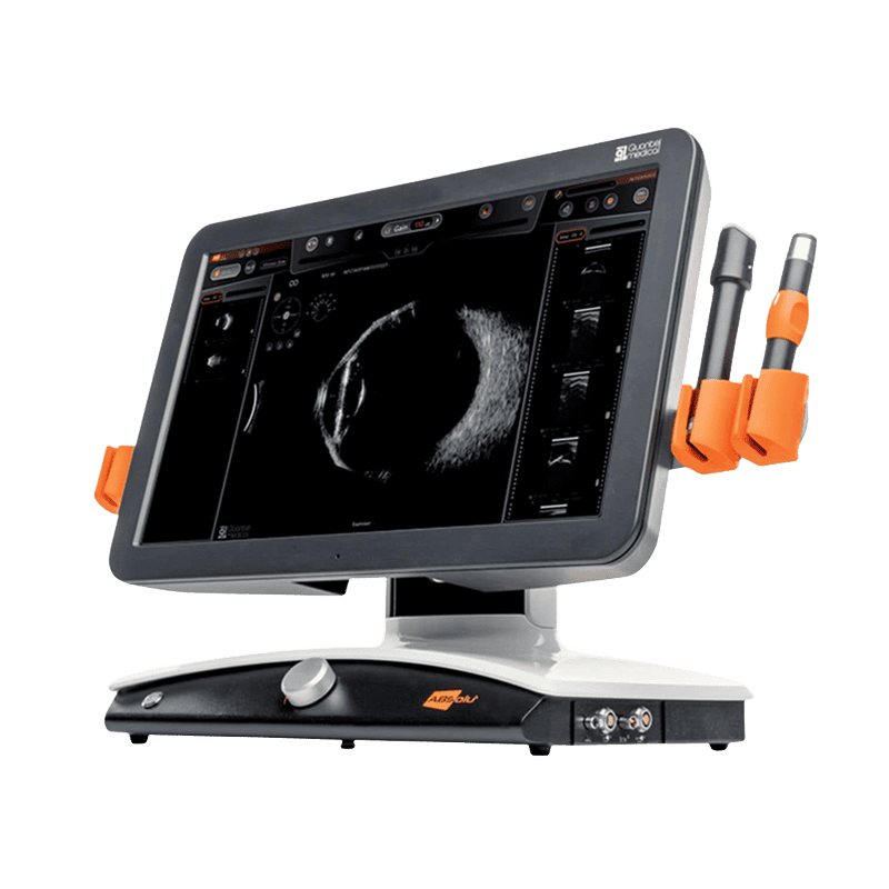
The Quantel Medical ABSolu® is a premium, all-in-one ultrasound platform that serves as the successor to the well-known Aviso system. It is widely considered one of the most advanced diagnostic ultrasound units in ophthalmology today, combining A-scan, B-scan, UBM, and Standardized S-mode into a single workstation. It is primarily used to determine suitable sizing for Implantable Collamer Lens (ICL) procedures and to assess the possibility of retinal pathologies such as retinal detachment, vitreous abnormalities, and other posterior segment conditions that may not be clearly visible on routine examination.
ELECTROPHYSIOLOGY AND IMAGING
Electrophysiology and Imaging uses advanced diagnostic technology to assess the health and function of your eyes in detail. These specialised tests help detect and monitor conditions affecting the retina, optic nerve, and visual pathways, often before noticeable vision changes occur. By combining precise imaging with functional eye testing, our eye specialists can make accurate diagnoses, personalise treatment plans, and monitor your progress to help protect your vision.
RETI-port/scan 21 - Beta Plus Advance
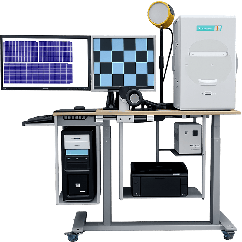
The RETI-port/scan 21 beta plus advanced system supports key electrophysiological tests such as ERG (Electroretinography), VEP (Visual Evoked Potentials), and EOG (Electrooculography), which measure how different parts of the visual system function electrically. ERG evaluates the retina’s response to light to detect retinal diseases, VEP assesses the visual pathway from the eye to the brain to identify neurological or optic nerve disorders, and EOG measures retinal pigment epithelium function to help diagnose conditions affecting retinal health and eye movement control.
RETI-port/scan 21 - Beta Plus Advance
The RETI-port/scan 21 beta plus advanced system supports key electrophysiological tests such as ERG (Electroretinography), VEP (Visual Evoked Potentials), and EOG (Electrooculography), which measure how different parts of the visual system function electrically. ERG evaluates the retina’s response to light to detect retinal diseases, VEP assesses the visual pathway from the eye to the brain to identify neurological or optic nerve disorders, and EOG measures retinal pigment epithelium function to help diagnose conditions affecting retinal health and eye movement control.
CONTRAST SENSITIVITY TEST
A contrast sensitivity test is a visual assessment that measures your ability to distinguish between an object and its background, particularly in low-contrast conditions or varying shades of gray. While a standard eye chart (like the Snellen chart with big black letters on a white background) measures visual acuity—how sharp or clear your vision is at a distance—contrast sensitivity evaluates how well you see in real-world environments, such as low light, fog, or glare.

Mars Contrast Sensitivity Test
The Mars Contrast Sensitivity Test (available in Letter versions) is a standardized, small-format clinical vision assessment used to evaluate peak visual contrast sensitivity. Unlike standard visual acuity tests that measure the ability to see fine detail at high contrast (black letters on a white background), the Mars test evaluates how effectively the visual system processes lower contrast levels. It is vital for detecting subtle vision degradation, glare sensitivity, and functional visual impairment caused by corneal irregularities, cataracts, glaucoma, or retinal conditions—even when a patient achieves 20/20 visual acuity on standard eye charts.
FREQUENTLY ASKED QUESTIONS
Cataract & Vision Correction Surgery
We use advanced Biometry tools like the ZEISS IOLMaster 700 and Topcon Aladdin. These machines use precise technology to measure through even dense cataracts, ensuring our surgeons can perfectly select your standard, premium, or Toric intraocular lenses.
We utilize the Topcon SP-1P for Corneal Specular Microscopy. This automated device analyzes the innermost layer of your cornea (endothelial cells) to confirm your eye is healthy enough to undergo and recover from procedures like cataract surgery safely.
Safety is our priority. We use comprehensive 3D Corneal Topography and Tomography—including the OCULUS Pentacam, SCHWIND Sirius, and the highly advanced SCHWIND MS39—to map the entire structure of your eye and rule out risks like early keratoconus.
We use the ZEISS VISUMAX 800, a next-generation femtosecond laser that powers our SMILE® pro 2.0 procedures. Its ultra-fast frequency allows us to complete laser vision treatments safely in under 10 seconds.
Dry Eye Diagnosis & Treatment
We eliminate the guesswork using automated ocular surface analyzers like the IDRA and the Lumibird C-Diag. These non-invasive machines perform 3D meibography and tear film stability tests to pinpoint exactly what is irritating your eyes.
Yes. We offer the C-STIM (an Intense Pulsed Light or IPL system) and the E-Eye. Both are specialized, non-invasive treatments designed to neurologically stimulate your meibomian glands, restoring your natural oil production for long-lasting relief.
Retina & Advanced Diagnostics
In many cases, yes. We use the Optos California, an ultra-widefield imaging system that captures an expansive 200-degree view of your retina in a single, non-contact scan. This reveals hidden peripheral diseases, tears, or detachments that traditional narrow-angle cameras often miss.
Our ZEISS CIRRUS™ HD-OCT and Topcon Maestro2 use high-speed 3D imaging and AI analytics to track microscopic anatomical changes in your retina and optic nerve. This allows our specialists to detect and monitor sight-threatening conditions like macular degeneration as early as possible.
Yes. We use Ophthalmic Ultrasonography (eye ultrasound) and the Topcon DRI OCT Triton (Swept-Source OCT). These technologies use high-frequency sound waves or deep-penetrating 1,050nm wavelength lasers to safely see through dense cataracts and evaluate the back of your eye.
Glaucoma & Vision Mapping
We use a Visual Field Test to measure your entire scope of vision, including your central and peripheral (side) vision. It is a crucial diagnostic tool for detecting the subtle, progressive vision loss typically caused by glaucoma or other conditions affecting the optic nerve.
We evaluate this using a Contrast Sensitivity Test. While standard eye charts only check the smallest letters you can read in perfect lighting, a contrast test measures your ability to distinguish objects from their background. This is critical for diagnosing early glaucoma or cataracts and ensuring you can drive safely at night.
Specialized Muscle & Nerve Tests
We use a Hess Chart (or Hess Screen Test) to accurately map the movement and coordination of your extraocular muscles. This specialized diagnostic tool allows us to pinpoint exactly which eye muscles are underacting or overacting, which is essential for treating strabismus (squint) or diplopia (double vision).
Electrophysiology testing evaluates the actual electrical activity of your entire visual pathway—from the retina to the brain. We use this advanced diagnostic tool to accurately diagnose and monitor complex, inherited, or unexplained retinal diseases and optic nerve disorders that standard imaging cannot fully detect.
Operating Room Technology
OasisEye Specialists is the first eye centre in Malaysia to offer the ZEISS ARTEVO 750 iOCT, a next-generation digital microscope with real-time 3D OCT imaging overlays. We also use the premium Leica M822, providing our surgeons with world-class, high-definition views for micron-level precision.
We evaluate this using a Contrast Sensitivity Test. While standard eye charts only check the smallest letters you can read in perfect lighting, a contrast test measures your ability to distinguish objects from their background. This is critical for diagnosing early glaucoma or cataracts and ensuring you can drive safely at night.