
Reviewed by Dr Amir Samsudin, Glaucoma Specialist of OasisEye Specialists | Last updated: September 2026
Why Glaucoma Matters in Malaysia
Glaucoma is often called the “silent thief of sight” because it typically steals vision so gradually that most people do not notice any change until significant, permanent damage has occurred. According to the Malaysian National Eye Survey II (NES II), glaucoma is recognised as the second leading cause of irreversible blindness in Malaysia, accounting for approximately 6.6% of all blindness cases nationwide. Furthermore, global epidemiological projections published in Ophthalmology (Tham et al., 2014) estimate that over 76 million people worldwide live with glaucoma, a number expected to exceed 111 million by 2040 – with a disproportionately heavy burden falling on Asian populations.
Because fluid buildup damages the optic nerve fibres silently, understanding your personal risk and securing timely eye examinations are your primary defences against irreversible vision loss.
Causes and Risk Factors
Glaucoma develops when fluid (aqueous humor) inside the front portion of the eye cannot drain properly through its natural drainage channels. This obstruction increases fluid buildup and raises internal eye pressure, gradually squeezing and killing the delicate nerve fibres that transmit visual signals to your brain.
Key risk factors include:
- Age over 40 years: As the body ages, the eye’s natural fluid drainage channels become less efficient, increasing internal pressure risk; data from the Ministry of Health Malaysia Clinical Practice Guidelines (MOH CPG 2017) note that glaucoma prevalence rises exponentially after age 40.
- Family history of glaucoma: Genetic variations inherited from relatives can alter the structural development or resistance of your eye’s drainage system, leaving you up to four times more likely to develop the condition.
- Ethnicity: People of East Asian and Southeast Asian descent have a higher structural predisposition to narrow drainage angles (leading to “angle-closure glaucoma”), whereas individuals of African or South Asian heritage face significantly higher rates of “open-angle glaucoma.”
- Elevated internal eye pressure (Intraocular Pressure/ IOP): Higher fluid pressure directly compresses the optical nerve head, impairing vital blood flow to delicate nerve cells.
- High myopia (severe short-sightedness) or hyperopia (far-sightedness): Anatomical stretching of the eyeball in high myopia weakens the optic nerve tissue, while smaller, compact eyeballs in hyperopia physically crowd the drainage angle.
- Prolonged use of steroid medications: Steroid eye drops, oral tablets, or creams can alter the cellular structure of the eye’s drainage meshwork, causing fluid pressure to spike.
- Pre-existing medical conditions: Chronic conditions like diabetes and high blood pressure disrupt blood circulation to the optic nerve, reducing its ability to withstand normal internal eye pressure.
- Previous eye injury or surgery: Physical trauma can cause internal scarring that permanently blocks drainage pathways months or years after the original event.
Stages of Glaucoma: What They Mean for You
Glaucoma is categorized by structural damage to the nerve and functional visual loss:
| Stage | What is Happening Inside the Eye | Practical Impact on Daily Life |
|---|---|---|
| Early/ Mild | Microscopic loss of nerve fibres; internal pressure may be elevated or normal. | No noticeable visual symptoms. You can see clearly, drive, and read without difficulty. Only detailed clinical imaging can detect this stage. |
| Moderate | Noticeable thinning of the optic nerve head; microscopic dead spots form in peripheral (side) vision. | Side vision begins to dim, though central vision remains sharp. Most patients still do not notice these gaps because the brain automatically “fills in” the missing areas. |
| Advanced | Severe damage to the optic nerve bundle; significant loss of side vision. | “Tunnel vision” occurs. Navigating stairs, driving, or noticing objects to your side becomes difficult. High risk of tripping or bumping into objects. |
| End-Stage | Extreme optic nerve destruction; only a narrow island of central vision or light perception remains. | Severe visual impairment. Reading and recognizing faces become extremely challenging or impossible without specialized low-vision aids. |
Note on Outcomes: Vision already lost to glaucoma cannot be restored by any current medical or surgical treatment. However, early detection and consistent management are associated with a significantly reduced rate of disease progression, helping the majority of patients maintain useful functional vision for the rest of their lives.
Signs and Symptoms
Glaucoma manifests differently depending on whether it develops slowly over years (Open-Angle Glaucoma) or rapidly within hours (Acute Angle-Closure Glaucoma).
What Only a Clinician Can Detect During an Exam
- Elevated intraocular pressure (measured using a tonometer).
- Structural cupping or thinning of the optic nerve (observed during a dilated eye exam).
- Microscopic loss of peripheral visual fields (detected via automated visual field testing).
- Narrow or blocked drainage angles inside the front of the eye (evaluated via gonioscopy).
What Patient or Family Members Might Notice at Home
- In Chronic Glaucoma (Open-Angle):
- Subtle loss of side (peripheral) vision (often unnoticed until advanced).
- Difficulty navigating in dim or low-light conditions.
- In Acute Glaucoma (Acute Angle-Closure – A Medical Emergency):
- Sudden, severe pain in or around the eye.
- Severe headache often accompanied by nausea or vomiting.
- Rapidly blurred vision or sudden loss of sight.
- Seeing rainbow-coloured circles or “halos” around bright lights.
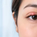
- Redness in the eye accompanied by a hard, firm feeling when gently touched through the eyelid.
Urgent Warning: If you, your parent, or a family member experience sudden eye pain, red eyes, vision blurring, and nausea, seek immediate care at an emergency room or specialised eye clinic. Acute angle-closure glaucoma requires urgent medical pressure-lowering treatment within hours to preserve sight.
Treatment Options
Treatment for glaucoma focuses on one core objective: lowering internal eye pressure to prevent further damage to the optic nerve.
- Prescription Eye Drops (Medication)
- What it is: Daily medicated drops (such as prostaglandin analogues, beta-blockers, or alpha-agonists) that either reduce the amount of fluid the eye produces or improve fluid drainage.
- What it is for: Primary first-line treatment for mild to moderate chronic glaucoma.
- Limitations & Risks: Eye drops do not cure glaucoma; they only control fluid pressure while actively used. They require lifelong daily compliance. Side effects may include eye redness, eyelashes lengthening, stinging, or system-wide effects (e.g. changes in heart rate or asthma exacerbation depending on the medication class).
- Laser Therapy (e.g., SLT or LPI)
- What it is: In-office laser procedures. Selective Laser Trabeculoplasty (SLT) uses gentle light energy to stimulate drainage tissue in open-angle glaucoma. Laser Peripheral Iridotomy (LPI) creates a microscopic relief hole in the iris for narrow-angle glaucoma.
- What it is for: Used as a primary or supplementary treatment to lower eye pressure without relying solely on drops.
- Limitations & Risks: The pressure-lowering effect of laser treatments is typically temporary, often wearing off after 1 to 5 years, though procedures like SLT can sometimes be repeated. Transient pressure spikes or mild inflammation may occur post-procedure.
- Surgical Procedures (Trabeculectomy & Glaucoma Drainage Implants)
- What it is: In-operating-theatre surgery where a microscopic drainage channel (filtering bleb) or tiny tube shunt is created to bypass natural drainage pathways and drain fluid under the outer lining of the eye.
- What it is for: Advanced glaucoma or cases where medication and laser therapy fail to lower eye pressure sufficiently.
- Limitations & Risks: Surgery carries higher procedural risks, including infection (endophthalmitis), abnormally low eye pressure (hypotony), cataract formation, or scarring that closes the surgical flap over time, potentially requiring revision surgery.
- Minimally Invasive Glaucoma Surgery (MIGS)
- What it is: Modern, microscopic implants or tiny micro-stents inserted into the eye’s drainage channel, often performed alongside routine cataract surgery.
- What it is for: Mild to moderate glaucoma requiring modest pressure reduction with a faster recovery profile.
- Limitations & Risks: MIGS generally offers a milder pressure drop compared to traditional major surgery (like trabeculectomy) and may not be suitable for patients with severe, advanced glaucoma.
Screening and Prevention
Because chronic glaucoma produces no pain and no visible symptoms in its early stages, regular clinical screening is the only reliable way to detect the condition before vision is permanently impaired.
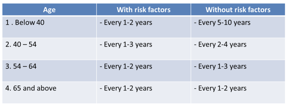
The Ministry of Health Malaysia CPG (2017) and the American Academy of Ophthalmology (AAO) Preferred Practice Pattern (2020) recommend the following baseline screening schedule:
- Adults aged 40 to 54: Comprehensive eye exam every 2 to 4 years.
- Adults aged 55 to 64: Comprehensive eye exam every 1 to 3 years.
- Adults aged 65 and older: Comprehensive eye exam every 1 to 2 years.
- Individuals with high risk factors (family history of glaucoma, extreme myopia, long-term steroid use): Annual comprehensive eye exams starting at age 40 (or earlier as advised by an eye specialist).
A proper glaucoma screening must include more than a simple eye chart reading. It requires tonometry (measuring eye pressure), gonioscopy (checking the drainage angle), funduscopy/OCT imaging (examining the optic nerve head), and visual field testing (checking peripheral vision).





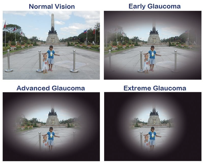 What makes glaucoma more dangerous is that the most common type of glaucoma does not show any obvious symptoms, hence goes undetected in many people. Thus, glaucoma is known as the “