
Reviewed by Dr Rawindar Singh Ranu A/L Harchand Singh, Refractive Surgeon of OasisEye Specialists | Last updated: August 2026
What Is Presbyopia?
Presbyopia is the gradual, age-related loss of the eye’s natural ability to focus on nearby objects.
Presbyopia (often referred to locally as rabun tua) is an inevitable part of the human aging process that typically becomes noticeable around the age of 40. It affects nearly 100% of the population by their mid-to-late 40s. While it can be frustrating to find yourself holding menus at arm’s length or constantly searching for reading glasses, it is not a disease—it is a physiological shift in how the eye focuses.
Causes and Risk Factors
Inside a young eye, the natural lens is soft, flexible, and changes shape effortlessly to switch focus between far and near objects. As time passes, two main changes occur:
- Stiffening of the Natural Lens: The clear crystalline lens inside your eye gradually hardens and loses its elasticity over time, making it physically harder for the surrounding focusing muscles to squeeze the lens into a shape that bends near light clearly.
- Natural Cellular Aging: As proteins within the eye’s lens slowly change structure with age, the lens loses its mechanical flexibility regardless of whether you previously had perfect 20/20 vision, near-sightedness (myopia), farsightedness (hyperopia), or astigmatism.
- Extended Digital Micro-Focusing: While screen time does not cause presbyopia, modern reliance on smartphones and laptops increases the visual strain caused by declining near focus, causing symptoms to be noticed earlier in everyday life.
Staging and Practical Progression
Presbyopia develops along a continuum. What you experience in daily life shifts as the natural lens continues its natural hardening process:
- Early Onset (Ages 40–44): You may notice slight eye strain or delay when switching focus from a smartphone to a far wall, particularly in low-light environments like dimly lit restaurants.
- Established Presbyopia (Ages 45–52): Reading fine print without dedicated reading glasses or progressive lenses becomes consistently difficult. Arm-length holding of text becomes a daily habit.
- Advanced Presbyopia (Ages 53+): Near vision without optical correction is significantly blurred across all standard reading distances, and frequent prescription updates for reading glasses are required every few years.
Signs and Symptoms
Because presbyopia progresses gradually, understanding what you can monitor at home versus what requires clinical evaluation helps you seek timely care.
What You Might Notice at Home
- Needing to hold books, smartphones, or food labels further away to see them clearly.
- Frequent eye strain, visual fatigue, or mild frontal headaches after prolonged near work or computer reading.
- Requiring brighter, direct lighting to read print that used to be clear in ambient light.
- Blurred vision when switching your gaze quickly between close-up tasks and distance viewing.
What Only an Eye Care Clinician Can Detect
- Loss of Accommodation Amplitude: Micro-measurements revealing the exact loss of mechanical focusing power in diopters.
- Corneal and Lens Aberrations: Advanced topographic and wavefront scanning measuring how light rays bend through your unique cornea and lens.
- Early Cataract Formation: Differentiating between standard natural lens stiffening and early lens clouding (cataract development).
Treatment Options: Understanding Presbyond 2.0
To treat presbyopia, optical correction must restore functional focus across near, intermediate, and far distances.
| Feature | Presbyond 2.0 Laser Blended Vision | Traditional Monovision (Laser/ Implantable Lenses) | Refractive Lens Exchange (RLE)/ Premium IOLs |
|---|---|---|---|
| Surgical Technique | Wavefront-guided corneal laser shaping (ZEISS MEL 90) extending depth of field in both eyes | Laser or contact lens correction setting one eye strictly for distance, one strictly for near | Intraocular surgery removing the natural lens and replacing it with an artificial multifocal/EDOF IOL |
| Intermediate Vision & Blend Zone | Yes — Creates a continuous “Blend Zone” for seamless intermediate (computer screen) focus | No — Leaves a distinct focal gap in the middle distance, causing blurry intermediate vision | Variable — Focus depends on specific intraocular lens optics (multifocal vs. EDOF) |
| Ideal Candidate | Patients aged 40+ seeking independence from reading glasses, bifocals, or progressives | Patients seeking basic, entry-level correction for distance and near reading | Patients with severe presbyopia, hyperopia, or early natural lens changes |
| Patient Adaptation / Tolerance Rate | Up to 97% successful neuroadaptation rate (ZEISS published data) | 59% – 67% adaptation rate due to loss of stereo depth perception (published clinical data) | Dependent on patient adaptation to multifocal optical profiles |
| Primary Limitations & Risk Profile | Corneal surface procedure; natural aging continues, with a long-term retreatment rate of 4.17% – 5.81% (Current Eye Research) | Reduced stereoscopic depth perception and poor intermediate desktop vision | Invasiveness of intraocular surgery; potential for night-time glare or light halos around headlights |
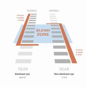
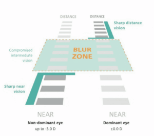
Image: Presbyond 2.0’s Blend Zone vs. Conventional Monovision’s Blur Zone in Intermediate Vision
Screening and Candidacy Evaluation
Presbyond 2.0 is highly effective, but thorough pre-operative screening is vital to determine whether corneal laser treatment is right for you.
Your specialist will determine your eligibility based on current clinical guidelines, detailed corneal diagnostic scans, and clinical health protocols. Standard candidacy assessments evaluate:
- Corneal thickness, structural integrity, and topography (mapping corneal shape).
- Tear film quality and dry eye health.
- Complete retinal and macula evaluation to ensure the back of the eye is fully healthy.
- Binocular vision testing and trial tolerance to ensure your brain comfortably adapts to the Blend Zone.