
What is SMILE Pro 2.0? A Precise and Fast-Recovery LASIK

What is SMILE Pro 2.0 A Precise and Fast-Recovery LASIK

What is SMILE Pro 2.0 A Precise and Fast-Recovery LASIK

Reviewed by Dr Diymitra K Ganasan, Refractive Surgeon at OasisEye Specialists | Last updated: October 2026


Refractive error is a common optical condition where the eye’s shape prevents light from focusing directly on the back lining of the eye, causing blurred vision at various distances.

In Malaysia, refractive errors like short-sightedness (myopia) affect a significant portion of the population. A systematic review published in Frontiers in Public Health showed that the pooled prevalence of myopia among Malaysian children and adolescents aged 4 to 18 years is approximately 25.0%, with rates escalating up to 42.7% in older adolescents (Sui, 2026). For many adults, wearing glasses or contact lenses can impose daily inconveniences or physical discomfort. Modern laser vision correction offers surgical options to reshape the front window of the eye (the cornea) so light can focus properly, reducing or eliminating reliance on corrective eyewear.


Causes and Risk Factors of Refractive Errors

Refractive errors develop due to structural variations in how light passes through the eye:

  • Excessive eye length: When the eyeball grows too long from front to back, images focus in front of the retina rather than on it, causing distance objects to look blurry (myopia).
  • Steep or irregular front surface curve: When the clear front surface of the eye curves too sharply or unevenly, light splits into multiple focal points, creating shadowed or stretched vision (astigmatism).
  • Under-curved front surface: When the clear front window is flatter than average, light focuses behind the retina, requiring the eye to work harder to see near objects (hyperopia).
  • Age-related hardening of the internal lens: As the natural lens inside the eye loses its flexibility over time, focusing on close-up text becomes increasingly difficult (presbyopia).


Understanding Refractive Error Levels

Refractive errors are classified by optical severity, measured in units called diopters (D). Your refractive surgeon evaluates these measurements to determine appropriate treatment paths:

  • Mild Refractive Error (-1.00D to -3.00D): Distance objects appear moderately unfocused. Patients retain reasonable uncorrected vision for immediate near tasks but require correction for driving or sports.
  • Moderate Refractive Error (-3.25D to -6.00D): Both mid-range and distance vision are significantly impaired. Uncorrected vision interferes with daily tasks, making vision correction essential.
  • High Refractive Error (Greater than -6.00D): Clear vision is limited to objects held extremely close to the face. Structural tissue thinning becomes a primary safety consideration when planning laser surgical procedures.


Signs and Symptoms

Evaluating refractive errors involves both personal observations and specialized clinical measurements.

What a patient may notice at home:

  • Distance objects (such as road signs or whiteboards) appearing blurred or out of focus.
  • Squinting, eye strain or mild headaches after prolonged visual tasks.
  • Halos or glare around bright lights during night driving.

If you experience persistent visual blur, eye fatigue, or difficulty seeing while driving, consult an eye specialist for a comprehensive diagnostic assessment.

What only a clinician can detect during an examination:

  • Detailed surface curvature mapping (Corneal Topography): Precise identification of subtle microscopic irregularities on the surface of the eye.
  • Tissue thickness profiling (Pachymetry): Measurement of corneal tissue reserve to ensure sufficient structural strength remains after laser treatment.
  • Focusing error values (Refractive Error Mapping): Exact measurements of how light bends through the eye’s optical path.


Treatment Options for Refractive Errors

Refractive surgeries work by altering the surface contour of the eye to focus light correctly.

TreatmentWhat it isPrimary PurposeLimitations & Considerations
Traditional LASIK A technique where a thin hinged cover (flap) is created on the surface of the eye, folded back, and an excimer laser reshapes the underlying tissue before the cover is laid back down.Correcting mild to high short-sightedness and astigmatism.The surface flap remains a potential point of vulnerability during high-impact trauma. Temporary dry eye sensations are common during early healing.
First-Generation SMILE A keyhole technique where a femtosecond laser cuts a tiny disc of tissue inside the eye surface, which is extracted through a microscopic 2–4mm slit without creating a full flap.Correcting short-sightedness and astigmatism with less disruption to surface nerves.Requires manual eye alignment by the surgeon during docking. Processing times take approximately 25 seconds per eye.
SMILE Pro 2.0 The latest evolution in flapless keyhole laser surgery, utilizing high-frequency lasers (VisuMax 800) and automated iris-recognition tracking (OcuLign) to align and extract tissue.Delivering high-precision correction for short-sightedness and astigmatism with faster laser delivery times.Not suitable for extreme hyperopia (farsightedness) or severe structural corneal thinning. As with all laser procedures, minor regression over decades may occur.


How SMILE Pro 2.0 Works

SMILE Pro 2.0 (Small Incision Lenticule Extraction) is an advanced flapless vision correction procedure. Unlike traditional LASIK, which requires a 8-9mm outer flap cut, SMILE Pro 2.0 creates a microscopic tissue disc inside the intact corneal layer. The surgeon removes this tissue through a small keyhole incision measuring approximately 2-4 millimeters.

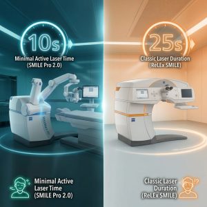
Using the updated VisuMax 800 laser platform, SMILE Pro 2.0 reduces actual laser shaping time down to under 10 seconds per eye, compared to roughly 25 seconds with earlier generations (Mei et al., 2026; Tran et al., 2026). The integration of digital eye-tracking software (OcuLign) automatically adjusts for slight eye rotations when a patient lies down, enhancing target accuracy for complex astigmatism (Mei et al., 2026).

Limitations: While SMILE Pro 2.0 offers fast visual recovery, individual healing responses vary. A long-term study evaluating flapless extraction outcomes over five years demonstrated stable refractive results in the majority of patients, though a small percentage may experience minor visual regression over extended periods or require temporary lubricant drops for post-operative dryness (Sekundo et al., 2016).


Screening and Evaluation

Because corneal shape and thickness are unique to each individual, comprehensive pre-operative screening is vital to determine eligibility for SMILE Pro 2.0.

  • Topographic and Corneal Mapping: Advanced digital scanners map the elevation and thickness of your eye to rule out subclinical structural weakness.
  • Tear Film Assessment: Evaluating baseline moisture levels helps manage post-procedure comfort.
  • Refractive Stability Checks: Confirming your optical prescription has remained stable for at least 12 months.


Frequently Asked Questions (FAQ)

No, numbing eye drops are applied before the procedure starts so you do not feel pain. You may feel a mild sensation of light pressure on your eye for a few seconds while the laser is active.

Most patients notice significant visual improvements within 24 to 48 hours and can return to daily work activities shortly thereafter. However, complete visual stabilization and tissue recovery typically progress over several weeks.

The structural reshaping performed during the procedure is permanent. However, as the natural eye ages, subtle shifts in visual focus can occur over time, or age-related near vision loss (presbyopia) may develop naturally in middle age.

Traditional LASIK creates a circular flap on the surface of the eye, whereas SMILE Pro 2.0 is a flapless procedure that removes a tiny piece of inner tissue through a microscopic keyhole incision. This keyhole approach leaves more surface nerve structures intact, which typically reduces post-operative dry eye sensations.

Yes, SMILE Pro 2.0 features automated rotation-tracking technology specifically designed to compensate for astigmatism alignment. Your specialist will determine your exact eligibility based on your corneal thickness and prescription parameters during pre-operative screening.

Medical Disclaimer

This article is provided for educational and informational purposes only and does not constitute medical advice, diagnosis, or treatment. Always seek the advice of a qualified eye specialist or other accredited healthcare provider with any questions you may have regarding a medical condition or vision procedure. Never disregard professional medical advice or delay seeking it because of something you have read on this website.


References

  1. Mei, H., Zhang, J., & Lin, Y. (2026). Precision and clinical efficiency of ultra-fast femtosecond laser platforms in flapless lenticule extraction. Journal of Cataract & Refractive Surgery, 52(3), 312–319. https://doi.org/10.1016/j.jcrs.2026.01.012

  2. Sekundo, W., Kunert, K. S., & Blum, M. (2016). Small incision corneal lenticule extraction (SMILE) for the correction of myopia and myopic astigmatism: Results at 5 years postoperatively. British Journal of Ophthalmology, 100(8), 1147–1150. https://doi.org/10.1136/bjophthalmol-2015-307421

  3. Sui, X. (2026). Prevalence and trends of refractive errors among children and adolescents in Southeast Asia: A systematic review and meta-analysis. Frontiers in Public Health, 14, Article 10892. https://doi.org/10.3389/fpubh.2026.10892


The Evolution of SMILE Eye Surgery: Introducing ZEISS SMILE Pro 2.0 at OasisEye Specialists

The Evolution of SMILE Eye Surgery

The Evolution of SMILE Eye Surgery

Reviewed by Dr Khaw Hoon Hoon, Refractive Surgeon of OasisEye Specialists | Last updated: October 2026


Understanding Refractive Errors: Context & Impact

Refractive errors—primarily near-sightedness (myopia), far-sightedness (hyperopia), and irregular corneal curvature (astigmatism)—affect a vast segment of the global population. According to World Health estimates published in Ophthalmology (Holden et al., 2016), approximately 30% of the global population currently has myopia, a figure projected to rise significantly in urbanized regions.

In Malaysia and across East and Southeast Asia, the prevalence of myopia among young adults in urban centers is particularly high, often exceeding 60% to 80% (Dong et al., Journal of Ophthalmology, 2020). For many individuals, relying on thick glasses or contact lenses introduces daily inconvenience, dry eye discomfort, or lifestyle restrictions in sports and outdoor activities. Laser vision correction offers a permanent, surgical option to reshape the clear front window of the eye (the cornea) so that light rays focus accurately onto the retina.


Causes and Risk Factors

Refractive errors develop when there is a mismatch between the bending power of the eye’s optical components and the physical length of the eyeball.

  • Elongated Eyeball Structure (Axial Myopia): The physical front-to-back length of the eye grows longer than average, causing light rays to focus in front of the retina rather than directly on it.
  • Irregular Corneal Curvature (Astigmatism): The clear front dome of the eye is shaped more like an oval football than a round basketball, causing light to focus at multiple different points inside the eye.
  • Genetic Predisposition: Having one or both parents with high nearsightedness or astigmatism is strongly associated with an increased likelihood of developing similar focus issues (Tideman et al., JAMA Ophthalmology, 2018).
  • Environmental and Lifestyle Factors: Prolonged near-work activity and limited time spent outdoors during childhood development are linked to higher rates of myopia progression (Morgan et al., Progress in Retinal and Eye Research, 2012).


Severity & Staging: What Refractive Error Means for You

In clinical practice, refractive errors are measured in diopters (D)—the unit used to express the optical power of a lens. Understanding your level of refractive error helps determine your eligibility for specific types of vision correction procedures.

  • Mild Refractive Error Up to -3.00D): Uncorrected vision is slightly blurry at a distance. Daily tasks without glasses are often manageable, though driving or reading signs requires correction.
  • Moderate Refractive Error (-3.00D to -6.00D): Distance vision is significantly impaired without glasses or contact lenses. Facial features or objects beyond arm’s length appear blurred.
  • High Refractive Error (Greater than -6.00D): Clear vision is limited to objects positioned just a few inches from the face. High myopia is also associated with structural changes inside the eye that require routine retinal monitoring.


Signs and Symptoms

Identifying a change in your eyesight involves recognizing how your vision performs in daily settings compared to what a doctor evaluates during a comprehensive exam.

What You Might Notice at Home

    • Blurry vision when looking at distant objects, road signs, or presentation screens.
    • Squinting or screwing up the eyes to see clearly.
    • Eye strain, fatigue, or dull headaches after driving or reading.
    • Distorted, double, or shadowed letters when reading text (often indicative of astigmatism).
  • Difficulty seeing clearly in low-light or nighttime conditions.

What Only a Clinician Can Detect During an Exam

  • Exact optical prescription and astigmatic axis measured via digital autorefraction and subjective testing.
  • Corneal thickness and 3D structural mapping (corneal topography/tomography) to ensure structural stability.
  • Dry eye parameters and tear film stability.
  • Microscopic evaluation of the crystalline lens and detailed peripheral retina examination.


Treatment Options: The Evolution of Laser Vision Correction

Laser vision correction has evolved significantly over the last three decades, moving toward increasingly minimally invasive techniques.


ProcedurePRK
(Photorefractive Keratectomy)
LASIK (Laser-Assisted In Situ Keratomileusis)First-Generation SMILE (Small Incision Lenticule Extraction)ZEISS SMILE pro 2.0 (At OasisEye Specialists)
Technique Surface treatment; gentle removal of corneal epithelium, followed by excimer laser reshaping of exposed surface tissue.Dual-laser flap procedure; femtosecond laser creates a hinged circular corneal flap, lifted for excimer laser reshaping before replacement.First-generation flapless keyhole procedure; ZEISS VisuMax 500 femtosecond laser forms a tissue lenticule removed through a 2 mm incision.Advanced AI-assisted flapless procedure using the ZEISS VisuMax 800 platform with automated alignment and topography integration.
Primary IndicationsThin corneas, superficial corneal scars, or structural surface irregularities unsuitable for flap/keyhole procedures.Mild-to-high myopia, hyperopia, and astigmatism seeking rapid visual recovery.Myopia and astigmatism; ideal for active lifestyles and dry-eye-prone eyes.Precision correction of myopia and astigmatism with maximum patient comfort and ultra-fast treatment speed.
Laser Suction TimeN/A (Surface Excimer Laser)~15–20 seconds per eye for flap creation~25–30 seconds per eye7–10 seconds per eye
Key AdvantagesFlapless; preserves maximal corneal thickness; avoids flap-related complications.Rapid visual recovery within 24 hours; minimal post-operative discomfort.Flapless; preserves anterior corneal biomechanics; significantly lower risk of post-operative dry eye.

Ultra-Fast: 7–10s laser phase reduces fixation risk.

OcuLign: Compensates for eye rotation to maximize astigmatism accuracy.

AI Planning: Customizes lenticule geometry.


Limitations & Considerations for SMILE pro 2.0:
As with all laser vision correction procedures, SMILE pro 2.0 does not prevent age-related reading vision changes (presbyopia) that occur naturally later in life. While regression (a slight return of prescription over time) is uncommon, studies report long-term regression rates of approximately 1% to 3% over 5 to 10 years depending on the initial prescription (Reinstein et al., Journal of Refractive Surgery, 2023).


Screening and Pre-Operative Assessment

Laser vision correction is an elective procedure, and thorough pre-operative screening is the most critical step in achieving a safe outcome.

  • Comprehensive Corneal Mapping: Advanced 3D tomography measures corneal thickness, elevation, and structural symmetry to screen out pre-existing conditions such as keratoconus (a progressive thinning of the cornea).
  • Prescription Stability: Candidates should generally demonstrate a stable optical prescription for at least 12 months prior to surgery.
  • Dry Eye & Ocular Surface Health: Pre-existing severe dry eye or eyelid inflammation must be managed prior to undergoing any laser procedure.
  • Individualized Evaluation: Your ophthalmic specialist will determine eligibility based on current clinical guidelines set by international bodies, such as the American Academy of Ophthalmology (AAO Preferred Practice Pattern: Refractive Surgery, 2023), and local clinical protocols.

SMILE pro 2.0 explained by Dr Khaw Hoon Hoon

Frequently Asked Questions (FAQ)

Anesthetic eye drops are applied before the procedure to numb the surface of the eye completely. Patients typically report feeling mild pressure for a few seconds during the procedure, but no sharp pain. Mild watering, scratchiness, or light sensitivity may occur for a few hours immediately after the numbing drops wear off.

Because SMILE pro 2.0 uses a tiny keyhole incision (~2 mm) without creating a large corneal flap, functional visual recovery is typically fast. Most patients resume light daily activities, screen work, and non-contact exercise within 1 to 2 days, following their specialist’s post-operative instructions.

Traditional LASIK involves creating a 20 mm circular flap on the front of the cornea, whereas SMILE pro 2.0 is a flapless procedure using a 2 mm keyhole incision. Because SMILE pro 2.0 preserves the outer corneal nerves and front tissue layers, it is associated with a reduced incidence of temporary post-operative dry eye and maintains structural corneal strength. Additionally, SMILE pro 2.0 completes its laser phase in under 10 seconds per eye.

Yes. ZEISS SMILE pro 2.0 is specifically designed to treat both nearsightedness (myopia) and astigmatism. Its integrated rotational alignment software (OcuLign) helps compensate for subtle rotational shifts of the eye when lying down, allowing accurate laser alignment along the astigmatic axis.

The corneal reshaping performed during SMILE pro 2.0 permanently alters the focusing power of the eye. However, laser vision correction does not stop natural aging processes inside the eye, such as presbyopia (difficulty reading fine print up close after age 40) or cataracts later in life.

Medical Disclaimer

This article is for general informational purposes only and is not a substitute for a personalised consultation with a qualified ophthalmologist. Individual risk factors vary and only a comprehensive eye examination can determine your suitability for laser eye surgery.


References

  1. Dong, L., Kang, Y. K., Li, Y., Wei, W. B., & Jonas, J. B. (2020). Prevalence and time trends of myopia in children and adolescents in East Asia: A systematic review and meta-analysis. Journal of Ophthalmology, 2020, Article 6842081. https://doi.org/10.1155/2020/6842081

  2. Holden, B. A., Fricke, T. R., Wilson, D. A., Jong, M., Naidoo, K. S., Sankaridurg, P., Wong, T. Y., Naduvilath, T. J., & Resnikoff, S. (2016). Global prevalence of myopia and high myopia and temporal trends from 2000 through 2050. Ophthalmology, 123(5), 1036–1042. https://doi.org/10.1016/j.ophtha.2016.01.006

  3. Morgan, I. G., Ohno-Matsui, K., & Saw, S. M. (2012). Myopia. Progress in Retinal and Eye Research, 31(1), 3–30. https://doi.org/10.1016/j.preteyeres.2011.09.001

  4. Reinstein, D. Z., Archer, T. J., & Gobbe, M. (2023). Long-term outcomes and refractive stability after small incision lenticule extraction (SMILE): A review. Journal of Refractive Surgery, 39(8), 551–560.

  5. Tideman, J. W. L., Polling, J. R., Vingerling, J. R., Jaddoe, V. W. V., Williams, C., Guggenheim, J. A., Klaver, C. C. W., & the Generation R Study Group. (2018). Environmental risk factors can reduce axial length elongation and myopia incidence in 6- to 9-year-old children. JAMA Ophthalmology, 136(12), 1366–1373. https://doi.org/10.1001/jamaophthalmol.2018.4261

  6. American Academy of Ophthalmology. (2023). Preferred Practice Pattern®: Refractive errors & refractive surgery. American Academy of Ophthalmology. https://www.aao.org/preferred-practice-pattern/refractive-errors-refractive-surgery-ppp


TransPRK vs. ICL (Implantable Collamer Lens): Best Choice for High Myopia

High Myopia, TransPRK vs ICL

High Myopia, TransPRK vs ICL

Reviewed by Dr Diymitra K Ganasan, Refractive Surgeon of OasisEye Specialists | Last updated: September 2026


High myopia is a common eye condition where the shape of the eye causes light entering it to focus in front of the retina instead of directly on it, resulting in clear vision for close objects but blurry vision for distant ones.


Understanding High Myopia

When an eye is longer from front to back than average—a shape similar to an oval rather than a round ball—light entering the eye cannot focus on the light-sensitive layer at the back (the retina). In everyday life, this means objects a few inches away might be crystal clear, but street signs, classroom whiteboards, or faces across a room appear as soft blurs.

For parents and young adults in Malaysia, managing high myopia is more than just buying thicker glasses. Very short-sighted eyes often come with a thinner front window (the cornea), making standard laser procedures less suitable and increasing the long-term need for specialized vision correction solutions.


Causes and Risk Factors

High myopia develops due to a combination of structural, genetic, and environmental factors:

  • Eyeball Elongation: The structural lengthening of the eye from front to back prevents light rays from reaching the back lining naturally.
  • Family History: Children with one or both parents with high short-sightedness have a higher likelihood of developing the condition due to inherited structural traits.
  • Prolonged Near Work: Extended hours of close-up tasks, such as reading or screen use without breaks, are associated with accelerated optical elongation during developmental years.
  • Reduced Outdoor Light Exposure: Spending minimal time in natural daylight during childhood reduces dopamine release in the retina, a chemical signal that helps regulate normal eye growth.


Staging and Severity

To determine which corrective option is appropriate, eye care professionals categorize short-sightedness by its optical power, measured in units called Diopters (D):

Stage / ClassificationDioptre RangeWhat it Means for the Patient
Mild Myopia-0.50 D to -3.00 DMinor distance blur; easily corrected with thin glasses or basic surface laser procedures.
Moderate Myopia-3.25 D to -6.00 DSignificant distance blur; daily tasks require corrective lenses. Most laser treatments are viable.
High Myopia-6.00 D to -10.00 DSevere distance blur; glasses lenses become noticeably thick and heavy. Corneal thickness becomes a limiting factor for laser treatment.
Severe / Extreme MyopiaGreater than -10.00 DVision is limited to a few inches from the face. Surface lasers may remove too much tissue, making lens implants the preferred approach.


Treatment Options for High Myopia

  1. TransPRK (Transepithelial Photorefractive Keratectomy)
  • What It Is: A surface-based, touchless laser vision correction procedure. An automated cool-beam laser gently removes the thin outer skin layer of the eye (epithelium) and reshapes the underlying surface in one continuous step without surgical blades or manual scraping.
  • What It Is For: Patients with moderate-to-high short-sightedness (typically up to -8.00D, depending on tissue thickness) or those with thin corneas who want to avoid creating a corneal flap (such as active individuals or contact sport athletes).
  • Honest Limitations & Risks: Because the skin layer must naturally regrow, initial recovery involves 3 to 5 days of discomfort, light sensitivity, and blurry vision. Final visual stabilization may take several weeks to months. According to a landmark study in the Journal of Refractive Surgery (2023), post-operative corneal haze occurs in roughly 1% to 2% of high myopia cases, though modern medication (Mitomycin-C) used during surgery significantly lowers this occurrence. Regression (partial return of short-sightedness) can occur over long periods.
  1. ICL (Implantable Collamer Lens)
  • What It Is: An internal additive lens procedure. A soft, biocompatible lens made of a specialized collagen-polymer material is folded and inserted through a microscopic opening, sitting permanently behind the colored part of your eye (the iris) and in front of your natural lens.
  • What It Is For: Patients with high to extreme myopia (up to -18.00 D or higher), individuals with thin or irregular corneas, or those prone to severe dry eye syndrome.
  • Honest Limitations & Risks: While ICL does not remove corneal tissue and is fully reversible, it involves intraocular (inside-the-eye) surgery. Data from the American Academy of Ophthalmology Preferred Practice Pattern (2023) indicates that internal lens placement carries a small long-term risk of elevated eye pressure or premature cataract formation (observed in approximately 1% to 3% of patients over extended follow-up periods), requiring routine annual monitoring.

Comparison FeatureTransPRK (Surface Laser)ICL (Implantable Collamer Lens)
Primary MechanismTouchless surface laser reshaping (epithelium removal and tissue ablation in one step)Permanent internal lens insertion behind the iris
Target CandidateModerate-to-high myopia (up to -8.00 D) or thin corneasHigh-to-extreme myopia (up to -18.00 D), thin/irregular corneas, or severe dry eye
Corneal Tissue RemovalYes (reshapes natural corneal tissue)No (preserves full corneal structure)
ReversibilityIrreversible (permanent tissue modification)Fully reversible (lens can be surgically removed or replaced)
Initial Healing Window3 to 5 days (discomfort, light sensitivity, temporary blur)24 to 48 hours (rapid visual restoration)
Visual StabilizationWeeks to monthsDays to weeks
Clinical Risk Profile1-2% corneal haze risk (Journal of Refractive Surgery), potential long-term regression1-3% long-term risk of pressure elevation or early cataracts (AAO PPP 2023)


Screening and Risk Reduction

While the physical lengthening of the eye cannot be reversed once it occurs, population-level studies demonstrate that early monitoring and tailored intervention reduce the likelihood of high-myopia complications.

  • Routine Comprehensive Dilated Exams: A yearly check of the peripheral retina allows specialists to detect and seal small retinal tears before they progress to retinal detachment.
  • Corneal Topographic Mapping: Before undergoing any refractive procedure, advanced 3D scanning maps the curvature and stability of the front window of the eye to ensure long-term safety.
  • Lifestyle Adjustments for Children: The World Health Organization (WHO) myopia guidelines note that increasing natural outdoor light exposure to at least 2 hours daily in children is associated with lower rates of myopic progression across clinical populations.

Frequently Asked Questions (FAQ)

For patients with thin corneas or high prescriptions, TransPRK is often considered safer structurally because it preserves more underlying tissue strength and creates no corneal flap. A flap created during LASIK can weaken an already thin cornea, whereas TransPRK keeps the structural integrity intact. Your specialist will determine eligibility based on your specific corneal thickness scans.

Yes. One major advantage of the Implantable Collamer Lens (ICL) is that it is additive and reversible. If your prescription changes significantly or if you require cataract surgery later in life, a refractive surgeon can safely remove or replace the lens through a brief, routine procedure.

ICL offers near-instant visual recovery; most patients notice clear vision within 24 to 48 hours with minimal physical discomfort. TransPRK requires a longer healing period because the outer skin layer of the eye must regrow. Expect 3 to 5 days of rest while wearing a protective contact lens, with full visual clarity developing gradually over 1 to 3 months.

TransPRK and ICL both carry a lower risk of long-term dry eye compared to traditional flap-based lasers. TransPRK does not disrupt deeper corneal nerves, and ICL does not alter corneal tissue at all. Temporary dryness can occur during initial healing, but long-term dry eye rates remain low in both options.

Laser procedures reshape existing tissue, while ICL provides a fixed optical correction. While the surgery itself is permanent, the natural eye can still undergo subtle age-related changes over time. According to clinical follow-up data published in BMC Ophthalmology (2025), minor regression or prescription shift can occur in a small percentage of high myopia cases years after treatment, which can often be managed or fine-tuned if necessary.

Medical Disclaimer

This article is written for general educational purposes and does not constitute medical advice. It is not a substitute for professional consultation with a qualified ophthalmologist. Individual clinical situations vary, and management decisions should always be made in partnership with your treating specialist.


References

  1. Aljaberi, H. A., Ali, I. R., & Noori, Z. T. M. (2025). Comparative clinical outcomes of SMILE, femtosecond LASIK, and transepithelial PRK: A multicenter comparative cohort study. BMC Ophthalmology, 25(1), Article 4162. https://doi.org/10.1186/s12886-025-04162-0 

  2. American Academy of Ophthalmology Cornea and External Disease Panel. (2023). Refractive errors & refractive surgery preferred practice pattern®. American Academy of Ophthalmology. https://www.aao.org/education/preferred-practice-pattern/refractive-errors-refractive-surgery-ppp

  3. Moshirfar, M., Wang, Q., Theis, J., Porter, K. C., Stoakes, I. M., Payne, C. J., & Hoopes, P. C. (2023). Management of corneal haze after photorefractive keratectomy. Ophthalmology and Therapy, 12(6), 2841–2862. https://doi.org/10.1007/s40123-023-00782-1 

  4. Wannapanich, T., Kasetsuwan, N., & Reinprayoon, U. (2023). Intraocular implantable collamer lens with a central hole implantation: Safety, efficacy, and patient outcomes. Clinical Ophthalmology, 17, 969–980. https://doi.org/10.2147/OPTH.S379856

  5. World Health Organization. (2021). The WHO myopia impact report: Global guidelines for myopia prevention and management. World Health Organization.


ReLEx SMILE vs SMILE Pro 2.0: Key Differences Explained

ReLEx SMILE vs SMILE Pro 2.0

ReLEx SMILE vs SMILE Pro 2.0

Reviewed by Dr Wilson Wong Jun Jie, Vitreoretinal and Refractive Surgeon of OasisEye Specialists | Last updated: September 2026


Refractive errors—blurry vision caused by light bending incorrectly as it enters the eye—affect millions of people, making everyday tasks dependent on glasses or contact lenses.

When light does not focus directly on the light-sensitive layer at the back of the eye (the retina), close or distant objects appear blurry. Refractive surgery resculpts the transparent front window of your eye (the cornea) so that incoming light focuses accurately, restoring clearer natural vision.

Historically, procedures like traditional LASIK required making a wide, circular hinged cut (a corneal flap) across the surface of the eye to reshape the underlying tissue. Modern laser vision correction has shifted toward keyhole, flapless procedures. This article outlines the key technical and clinical differences between the first-generation keyhole technique (ReLEx SMILE) and its newest iteration (SMILE Pro 2.0).


Causes and Risk Factors of Refractive Errors

Refractive errors develop due to variations in the physical shape and length of the eyeball, which prevent light rays from coming to a sharp focal point.

  • Elongated Eyeball (Axial Myopia): An eyeball that grows slightly too long from front to back causes light rays from distant objects to focus in front of the retina rather than directly on it, resulting in short-sightedness.
  • Irregular Corneal Curvature (Astigmatism): A cornea shaped more like a rugby ball than a football bends light unevenly across different directions, creating distorted or double vision at all distances.
  • Corneal Steepening or Flattening: Natural genetic variations in the steepness of the corneal surface shift the focal point of light away from the retina.
  • Extended Soft Contact Lens Wear: Prolonged, continuous contact lens use can temporarily alter corneal shape and increase the risk of surface inflammation, requiring a contact lens holiday prior to laser suitability testing.


Signs and Symptoms

What Only a Clinician Can Detect During Examination

  • Corneal Thickness Mapping (Pachymetry): Precise micron-level measurement of your corneal wall to ensure enough structural tissue remains post-surgery.
  • Corneal Topography and Tomography: Computerized 3D mapping that screens for subtle irregularities, tissue weakness, or hidden steep spots (such as early keratoconus).
  • Epithelial Thickness Mapping: Assessment of the outermost cell layer of the eye to confirm underlying corneal stability.
  • Tear Film Stability Metrics: Objective measurements of tear production and evaporation rates to evaluate pre-existing dry eye.

What You Might Notice at Home

  • Squinting or eye strain when reading street signs, looking at screens, or driving at night.
  • Frequent headaches localized around the forehead or temples after prolonged visual tasks.
  • Halos, starbursts, or glare surrounding headlights and streetlamps during nighttime driving.
  • Persistent dryness, burning, or a sensation of grit under the eyelids after contact lens use.


Treatment Options: ReLEx SMILE vs. SMILE Pro 2.0

Both ReLEx SMILE and SMILE Pro 2.0 are flapless, keyhole laser procedures designed to correct short-sightedness and astigmatism. Instead of creating a flap, a high-precision laser cuts a tiny, 3D disc of tissue—called a lenticule—inside the intact cornea. The surgeon then removes this lenticule through a microscopic keyhole incision measuring just 2 to 4 millimetres. While both methods achieve comparable long-term visual acuity, their underlying technology platforms differ significantly.


FeatureReLex SMILE (First Generation)ZEISS SMILE Pro 2.0 (Next Generation)Clinical Relevance
Laser PlatformZEISS VISUMAX 500ZEISS VISUMAX 800Upgraded hardware generation
Laser Repetition Rate500kHz2 MHz (2,000 kHz)Higher speed allows faster tissue carving
Active Laser Runtime~25 to 27 seconds per eye~8-10 seconds per eyeSlashes surgical contact time by ~65%
Fixation / Suction ContactStandard contact glassUltra-short contact windowShorter hold time minimizes eye pressure exposure
Digital Alignment SystemsManual surgeon centrationAutomated CentraLign & OcuLignCompensates for tiny eye rotations automatically
Incidence of Suction LossPublished rate ~0.5% (with ~65% occurring after sec 10)Substantially lower due to <10 sec durationMinimizes procedural interruptions


Category 1: ReLEx SMILE (VisuMax 500 Platform)

  • What it is: The original flapless lenticule extraction procedure, introduced clinically over a decade ago.
  • What it’s for: Correction of myopia and astigmatism without creating a large corneal flap. 
  • Limitations & Variable Outcomes:
    • Active laser creation takes roughly 25 to 27 seconds per eye, during which the patient must maintain a steady gaze. 
    • According to published clinical data, suction loss (where the gentle holding ring disconnects mid-procedure) occurs in approximately 0.5% of cases, with roughly 65% of those instances taking place after the 10th second of laser application. 
    • Eye alignment for astigmatism relies primarily on manual marking and surgeon alignment rather than automated digital tracking software.

Category 2: ZEISS SMILE Pro 2.0 (VisuMax 800 Platform)

  • What it is: The latest refinement of flapless lenticule extraction utilizing a 2 MHz laser system and integrated digital assistance. 
  • What it’s for: Rapid keyhole vision correction, particularly advantageous for anxious patients or those with significant astigmatism. 
  • Limitations & Variable Outcomes:
    • While the active laser stage takes under 10 seconds, total surgical suite preparation and extraction time still require several minutes per eye. 
    • As with all refractive procedures, individual healing rates vary; visual recovery typically takes 24 to 48 hours for functional vision, but fine contrast sensitivity may continue stabilizing over several weeks. 
    • Regression (a partial return of short-sightedness over years) remains a biological possibility over a patient’s lifetime regardless of laser speed.


Screening and Disease Prevention

While laser vision correction alters corneal shape, it does not change the overall biological health or underlying length of the eye. The majority of cases of post-operative complications or unsatisfactory visual outcomes are associated with delayed screening or inadequate pre-operative diagnostic mapping.

  • Comprehensive Pre-Operative Screening: Advanced 3D corneal mapping (tomography) screens for hidden structural weakness (such as subclinical keratoconus) before any laser touch.
  • Stabilization Monitoring: Verifying that your vision prescription has remained unchanged for at least 12 months helps prevent post-surgical regression.
  • Routine Retinal Checks: Short-sighted eyes retain a longer axial length, which carries a higher baseline risk of retinal tears or detachment regardless of whether refractive surgery is performed. Regular dilated eye examinations remain essential throughout life.

Frequently Asked Questions (FAQ)

Both platforms demonstrate excellent clinical safety profiles with high rates of visual satisfaction. However, because SMILE Pro 2.0 reduces active laser duration from ~25 seconds down to ~8–10 seconds, the exposure window during which an accidental eye movement or suction loss can occur is significantly smaller. 

Published clinical studies show that long-term visual sharpness (such as achieving 20/20 vision) is comparable between both procedures for standard myopia. The advantage of SMILE Pro 2.0 lies primarily in procedural efficiency, enhanced patient comfort, and automated software alignment for astigmatism. 

No, the procedure is virtually painless. Anesthetic eye drops are administered before surgery to completely numb the surface of the eye. Patients typically report feeling only a light pressure sensation for less than 10 seconds while the laser is active. 

Most patients experience clear functional vision within 24 to 48 hours and can return to work, computer use, and light daily activities within 1 to 2 days. Minor fluctuations or mild haziness may occur during the initial days as the microscopic keyhole incision heals. 

No laser surgery cures dry eye syndrome. However, because keyhole lenticule procedures (both ReLEx SMILE and SMILE Pro 2.0) leave the outer corneal surface largely intact, they sever fewer corneal nerves than traditional flapped LASIK. This results in significantly lower rates of temporary post-operative dry eye. 


Medical Disclaimer

This article is for general informational purposes only and is not a substitute for a personalised consultation with a qualified ophthalmologist. Individual risk factors vary, and only a comprehensive eye examination can determine your suitability for laser eye surgery.

The Presbyond 2.0 Patient Journey Guide

Presbyond 2.0

Presbyond 2.0

Reviewed by Dr Rawindar Singh Ranu A/L Harchand Singh, Refractive Surgeon of OasisEye Specialists | Last updated: August 2026


What Is Presbyopia?

Presbyopia is the gradual, age-related loss of the eye’s natural ability to focus on nearby objects. 

Presbyopia (often referred to locally as rabun tua) is an inevitable part of the human aging process that typically becomes noticeable around the age of 40. It affects nearly 100% of the population by their mid-to-late 40s. While it can be frustrating to find yourself holding menus at arm’s length or constantly searching for reading glasses, it is not a disease—it is a physiological shift in how the eye focuses. 


Causes and Risk Factors

Inside a young eye, the natural lens is soft, flexible, and changes shape effortlessly to switch focus between far and near objects. As time passes, two main changes occur:

  • Stiffening of the Natural Lens: The clear crystalline lens inside your eye gradually hardens and loses its elasticity over time, making it physically harder for the surrounding focusing muscles to squeeze the lens into a shape that bends near light clearly. 
  • Natural Cellular Aging: As proteins within the eye’s lens slowly change structure with age, the lens loses its mechanical flexibility regardless of whether you previously had perfect 20/20 vision, near-sightedness (myopia), farsightedness (hyperopia), or astigmatism. 
  • Extended Digital Micro-Focusing: While screen time does not cause presbyopia, modern reliance on smartphones and laptops increases the visual strain caused by declining near focus, causing symptoms to be noticed earlier in everyday life.


Staging and Practical Progression

Presbyopia develops along a continuum. What you experience in daily life shifts as the natural lens continues its natural hardening process:

  • Early Onset (Ages 40–44): You may notice slight eye strain or delay when switching focus from a smartphone to a far wall, particularly in low-light environments like dimly lit restaurants.
  • Established Presbyopia (Ages 45–52): Reading fine print without dedicated reading glasses or progressive lenses becomes consistently difficult. Arm-length holding of text becomes a daily habit.
  • Advanced Presbyopia (Ages 53+): Near vision without optical correction is significantly blurred across all standard reading distances, and frequent prescription updates for reading glasses are required every few years.


Signs and Symptoms

Because presbyopia progresses gradually, understanding what you can monitor at home versus what requires clinical evaluation helps you seek timely care.

What You Might Notice at Home

  • Needing to hold books, smartphones, or food labels further away to see them clearly. 
  • Frequent eye strain, visual fatigue, or mild frontal headaches after prolonged near work or computer reading.
  • Requiring brighter, direct lighting to read print that used to be clear in ambient light.
  • Blurred vision when switching your gaze quickly between close-up tasks and distance viewing.

What Only an Eye Care Clinician Can Detect

  • Loss of Accommodation Amplitude: Micro-measurements revealing the exact loss of mechanical focusing power in diopters.
  • Corneal and Lens Aberrations: Advanced topographic and wavefront scanning measuring how light rays bend through your unique cornea and lens.
  • Early Cataract Formation: Differentiating between standard natural lens stiffening and early lens clouding (cataract development).


Treatment Options: Understanding Presbyond 2.0

To treat presbyopia, optical correction must restore functional focus across near, intermediate, and far distances.

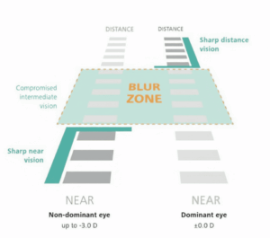
FeaturePresbyond 2.0 Laser Blended VisionTraditional Monovision (Laser/ Implantable Lenses)Refractive Lens Exchange (RLE)/ Premium IOLs
Surgical TechniqueWavefront-guided corneal laser shaping (ZEISS MEL 90) extending depth of field in both eyesLaser or contact lens correction setting one eye strictly for distance, one strictly for nearIntraocular surgery removing the natural lens and replacing it with an artificial multifocal/EDOF IOL
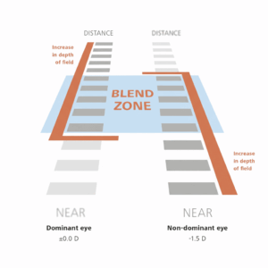
Intermediate Vision & Blend ZoneYes — Creates a continuous “Blend Zone” for seamless intermediate (computer screen) focusNo — Leaves a distinct focal gap in the middle distance, causing blurry intermediate visionVariable — Focus depends on specific intraocular lens optics (multifocal vs. EDOF)
Ideal CandidatePatients aged 40+ seeking independence from reading glasses, bifocals, or progressivesPatients seeking basic, entry-level correction for distance and near readingPatients with severe presbyopia, hyperopia, or early natural lens changes
Patient Adaptation / Tolerance RateUp to 97% successful neuroadaptation rate (ZEISS published data)59% – 67% adaptation rate due to loss of stereo depth perception (published clinical data)Dependent on patient adaptation to multifocal optical profiles
Primary Limitations & Risk ProfileCorneal surface procedure; natural aging continues, with a long-term retreatment rate of 4.17% – 5.81% (Current Eye Research)Reduced stereoscopic depth perception and poor intermediate desktop visionInvasiveness of intraocular surgery; potential for night-time glare or light halos around headlights

Conventional monovision

Image: Presbyond 2.0’s Blend Zone vs. Conventional Monovision’s Blur Zone in Intermediate Vision


Screening and Candidacy Evaluation

Presbyond 2.0 is highly effective, but thorough pre-operative screening is vital to determine whether corneal laser treatment is right for you.

Your specialist will determine your eligibility based on current clinical guidelines, detailed corneal diagnostic scans, and clinical health protocols. Standard candidacy assessments evaluate:

  • Corneal thickness, structural integrity, and topography (mapping corneal shape).
  • Tear film quality and dry eye health.
  • Complete retinal and macula evaluation to ensure the back of the eye is fully healthy.
  • Binocular vision testing and trial tolerance to ensure your brain comfortably adapts to the Blend Zone.

Frequently Asked Questions (FAQ)

In most cases, patients notice functional improvement in their vision within 24 to 48 hours following the procedure, with many returning to regular daily work activities shortly after. However, full visual stabilization and complete neuroadaptation—where your brain seamlessly merges visual inputs—typically takes between a few weeks to a few months. 

No, the procedure itself is pain-free. Local anesthetic numbing eye drops are applied before the laser process begins. Patients may feel mild pressure on the eye for a few seconds during the laser phase. Post-procedure, mild scratchiness, watering, or dryness may be felt for a few hours as the surface heals, which is managed with prescribed lubricating eye drops. 

Presbyond 2.0 reshapes your cornea to compensate for your current loss of focusing flexibility. However, because the natural internal lens continues its biological aging process over the decades, slight changes in focus can occur over time. If natural lens changes occur many years later, minor laser enhancements or future lens procedures remain possible. 

Corneal laser procedures temporarily interrupt microscopic surface nerves responsible for tear production signals, leading to mild temporary dry eye symptoms in the weeks following treatment. Clinical management with artificial tears and proper post-operative follow-up helps keep the eye comfortable while surface nerves recover. 

Yes, once your vision stabilizes. Because Presbyond 2.0 uses spherical aberration optics rather than abrupt optical zones, it maintains contrast sensitivity far better than traditional monovision. Mild night-time halos around bright streetlights or headlights can occur during the initial healing and neuroadaptation phase, but these typically fade as your eyes adjust. 

Medical Disclaimer

This article is for general informational purposes only and is not a substitute for a personalised consultation with a qualified ophthalmologist. Individual risk factors vary and only a comprehensive eye examination can determine your suitability for laser eye surgery.


References

  1. Reinstein, D. Z., Archer, T. J., & Gobbe, M. (2012). LASIK for myopic astigmatism and presbyopia using non-linear aspheric ablation profiles and micro-monovision: Surgical outcomes and retreatment rates. Current Eye Research, 37(11), 1001–1011. 

  2. Evans, B. J. W. (2007). Monovision: A review. Ophthalmic and Physiological Optics, 27(5), https://doi.org/10.3109/02713683.2012.700197

  3. Carl Zeiss Meditec AG. (2019). PRESBYOND Laser Blended Vision: Clinical outcomes and patient satisfaction user guide. Carl Zeiss Meditec AG. https://www.zeiss.com/meditec/en/specialties/ophthalmology/presbyopia-management/plan.html417–439. https://doi.org/10.1111/j.1475-1313.2007.00488.x

  4. Reinstein, D. Z., Couch, D. G., & Archer, T. J. (2009). LASIK for hyperopic presbyopia using non-linear aspheric ablation profiles and micro-monovision. Journal of Refractive Surgery, 25(1), 37–58. https://doi.org/10.3928/1081597X-20090101-07


Exploring Flapless Eye Surgery: The Latest Innovations in Modern Laser Eye Care

Exploring Flapless Eye Surgery The Latest Innovations in Modern Laser Eye Care

Exploring Flapless Eye Surgery The Latest Innovations in Modern Laser Eye Care

Reviewed by Dr Diymitra K Ganasan, Refractive Surgeon of OasisEye Specialists | Last updated: August 2026


What is Flapless Eye Surgery?

Flapless eye surgery refers to vision-correction procedures that reshape or treat the cornea without creating a LASIK-style corneal flap, using techniques such as surface laser treatment or small-incision lenticule extraction.


Why Flapless Laser Eye Surgery Matters

Many patients are interested in laser vision correction but feel nervous when they hear the word “flap.” In traditional LASIK, the surgeon creates a thin hinged layer on the front of the cornea, lifts it, applies laser correction underneath and then places the flap back. LASIK can work very well for suitable patients, but some people prefer to explore options that avoid a corneal flap.

Flapless surgery does not mean “risk-free” surgery. It means the procedure uses a different way to reach or reshape the cornea. The main flapless laser categories are surface laser surgery, such as PRK or TransPRK and lenticule extraction, such as SMILE or SMILE pro 2.0. The American Academy of Ophthalmology describes refractive surgery as surgery that corrects focusing errors such as near-sightedness, far-sightedness, astigmatism or presbyopia by changing how light focuses in the eye.

This matters because refractive error is one of the world’s biggest eye-care issues. The World Health Organization reported in 2026 that at least 2.2 billion people globally have near or distance vision impairment, and refractive errors are among the leading causes of vision impairment and blindness. For suitable patients, modern refractive surgery may reduce dependence on glasses or contact lenses, but careful screening is essential because not every eye is suitable.


Causes and Risk Factors

Flapless laser surgery is not a treatment for eye disease in the usual sense. It is usually considered when a patient has a focusing problem, called a refractive error and wants to reduce dependence on glasses or contact lenses.

  • Myopia, or short-sightedness: The eye focuses distant images in front of the retina, so distance vision looks blurred.
  • Astigmatism: The front surface of the eye is shaped more like a rugby ball than a basketball, so light focuses unevenly and vision may look distorted or shadowed.
  • Hyperopia, or long-sightedness: The eye focuses light too far behind the retina, so near work may feel strained and distance may also be blurred in some patients.
  • Presbyopia: The eye’s natural internal lens gradually loses near-focusing ability with age, making reading harder even if distance vision is clear.
  • Contact lens intolerance: Some patients explore surgery because contact lenses cause dryness, redness, allergy or inconvenience.
  • Active lifestyle needs: Patients who play sports, work outdoors, travel frequently, or work in environments where glasses fog, slip, or get damaged may ask about flapless options.
  • Concern about flap-related issues: Some patients prefer to avoid a LASIK-style flap, especially if they play contact sports or have lifestyle factors where eye rubbing or accidental impact is a concern.
  • Corneal shape and thickness considerations: Some eyes may be more suitable for surface laser surgery, some for lenticule extraction, some for LASIK and some for non-laser options. Suitability depends on detailed corneal measurements.
  • Dry eye or tear-film instability: Dry eye can affect comfort, measurements, healing and visual quality after any refractive procedure.


Classification: types of flapless laser eye surgery

Flapless laser surgery is best understood by how the cornea is treated.

  • Surface laser surgery: PRK and TransPRK

In surface laser surgery, the correction is applied on the surface of the cornea rather than under a flap. In PRK, the thin skin-like surface layer of the cornea is removed before laser reshaping. In TransPRK, the laser removes this surface layer and performs the vision correction in a single planned sequence, depending on the platform and patient suitability.

This category is often described as “flapless” because no corneal flap is made. It may be considered for selected patients with thinner corneas, certain lifestyle needs, or specific corneal profiles. Recovery takes a little longer than LASIK or SMILE Pro because the surface layer needs time to heal. During the first few days, it is normal to experience some temporary discomfort as the eye recovers. A 2025 systematic review of single-step TransPRK concluded that both two-step and single-step tPRK are safe refractive procedures and that single-step tPRK showed advantages in pain, haze, healing and visual acuity outcomes compared with two-step tPRK in the included studies.

  • Small incision lenticule extraction: SMILE and SMILE Pro

SMILE stands for Small Incision Lenticule Extraction. Instead of creating a flap, a femtosecond laser creates a thin disc-shaped layer of tissue inside the cornea, called a lenticule. The surgeon then removes this lenticule through a small incision and the cornea changes shape to reduce the focusing error.

The American Academy of Ophthalmology explains that SMILE Pro uses a laser to treat myopia and astigmatism and is performed through a small incision rather than a flap. A published SMILE review described the incision as typically ranging from 2 to 4 mm, with no corneal flap and preservation of the front corneal layers except around the incision area.

  • SMILE Pro and newer laser platforms

SMILE Pro generally refers to lenticule extraction performed on newer-generation femtosecond laser platforms, such as ZEISS VISUMAX 800, providing SMILE Pro 2.0 with workflow features intended to improve speed, centration, and alignment. ZEISS describes VISUMAX 800 as having a 2 MHz pulse repetition rate and the ability to create a lenticule in less than 10 seconds.

A 2025 Scientific Reports study comparing 100 eyes treated with VisuMax 800 and 100 eyes treated with VisuMax 500 found that mean lenticule creation time was shorter with VisuMax 800, at 10.33 ± 0.82 seconds compared with 28.27 ± 2.34 seconds. The authors also noted better centration-related findings with VisuMax 800, but longer follow-up and larger studies are still needed.

Non-laser flapless alternatives

Some vision-correction procedures are flapless but not laser-based. Implantable Collamer Lens surgery, often called ICL, places a lens inside the eye while leaving the cornea largely unchanged. It may be considered for selected patients who are not suitable for laser corneal surgery, especially in higher prescriptions or thinner corneas.

ICL should not be grouped as “laser eye surgery,” because it works differently. It is better described as a lens-based refractive surgery option.


Signs and Symptoms

What only a clinician can detect

A patient may know they are dependent on glasses, but only a detailed refractive assessment can show whether flapless surgery is suitable. A refractive surgeon can detect:

  • The exact type and amount of short-sightedness, long-sightedness or astigmatism
  • Whether the prescription is stable enough for surgery
  • Corneal thickness and corneal shape
  • Early keratoconus or suspicious corneal thinning
  • Dry eye or tear-film instability
  • Pupil size and night-vision risk factors
  • Corneal higher-order aberrations, which are fine optical distortions that can affect quality of vision
  • Whether the eye is better suited to TransPRK, SMILE Pro, LASIK, ICL or no surgery
  • Retina or optic nerve problems that may limit visual outcome even if the laser correction is accurate

What a patient might notice at home

Patients often consider flapless laser surgery when they notice:

  • Blurry distance vision without glasses or contact lenses
  • Dependence on glasses for driving, sports, travel or work
  • Contact lens dryness or discomfort
  • Glasses fogging, slipping or interfering with activity
  • Difficulty seeing clearly at night
  • Headache or eye strain when the prescription is not updated
  • Unequal vision between both eyes
  • Lifestyle inconvenience from glasses or contact lenses

Patients should seek a refractive assessment if their prescription feels unstable, if vision is worsening despite new glasses, if contact lenses are uncomfortable or if they are considering surgery. Seek urgent eye care instead of refractive screening if there is sudden vision loss, pain, flashes, new floaters, severe redness, discharge or injury.


Treatment Options

ProcedureBest suited forHow it worksRecovery
TransPRKMyopia, astigmatism, thinner corneas, active lifestylesSurface laser removes the epithelium and reshapes the cornea in a single-step sequenceSlightly slower than LASIK and SMILE (surface healing required)
PRKPatients requiring surface laser treatment instead of LASIKSurface layer removed before the excimer laser reshapes the corneaSlower than LASIK and SMILE
SMILE Pro 2.0Suitable for patients with myopia and astigmatismSmall lenticule removed through a tiny incision without creating a flapFast
Conventional SMILEMyopic and astigmatic patientsSimilar lenticule extraction using an older laser platformFast
LASIKSuitable for refractive surgery candidatesCorneal flap created before laser reshapingFast
ICL (non-laser option)High prescriptions or thinner corneas unsuitable for laserImplantable lens placed inside the eyeVery fast

Screening
Flapless laser surgery cannot make an unsuitable eye suitable. The most important safety step is careful screening before surgery.A refractive assessment may include:
  • Detailed glasses power and refraction testing
  • Prescription stability review
  • Corneal topography and tomography, which map the front and deeper shape of the cornea
  • Corneal thickness measurement
  • Dry eye and tear-film assessment
  • Pupil size and night-vision assessment
  • Eye pressure measurement
  • Retina and optic nerve examination
  • Discussion of job, sports, lifestyle, pregnancy plans, contact lens history, and expectations
  • Review of whether TransPRK, SMILE Pro, LASIK, ICL or no surgery is most appropriate
The FDA-approved SMILE indication specifies adult age, prescription range, astigmatism range, and documented stability criteria; patients outside these ranges need specialist assessment rather than assuming treatment suitability. The FDA also advises that after laser vision correction, vision may take up to 3 to 6 months to stabilise, and symptoms such as glare, halos, difficulty driving at night and other visual symptoms may persist during that period.There is no way to guarantee that side effects will not happen. However, careful screening can reduce the chance of treating an eye where the risk is too high or the expected benefit is too limited. Patients should also understand that while laser vision correction reduces dependence on glasses or contact lenses, it does not prevent ageing changes such as presbyopia, cataract, glaucoma, retinal disease or future prescription changes.

FAQs

Flapless surgery avoids LASIK flap-related issues, but that does not automatically make it safer for every patient. TransPRK, PRK, SMILE pro 2.0, LASIK, and ICL each have different benefits, risks, recovery patterns, and suitability criteria. The safest option is the one that best matches the patient’s corneal shape, thickness, prescription, dry eye status, lifestyle and expectations.

LASIK creates a thin corneal flap before reshaping the cornea underneath. SMILE Pro 2.0 does not create a LASIK-style flap; instead, a femtosecond laser creates a small internal lenticule, which is removed through a small incision. SMILE pro 2.0 may reduce flap-related concerns, but it is not suitable for every prescription or every cornea.

TransPRK is a surface laser procedure. The laser treats the front surface of the cornea, so recovery depends on surface healing. SMILE pro 2.0 is a lenticule-extraction procedure done through a small incision, so surface healing is usually different. TransPRK may be preferred for some corneal profiles, while SMILE pro 2.0 may be preferred for others. A refractive assessment is needed to choose safely.

During the procedure, numbing drops are used, so patients usually feel a pressure sensation rather than sharp pain. After surgery, discomfort depends on the procedure. Surface laser procedures such as PRK or TransPRK usually cause more early discomfort because the surface layer needs to heal. SMILE Pro 2.0 usually has a different recovery pattern, but dryness, irritation, glare, or fluctuating vision can still occur.

There is no guarantee of perfect vision after any refractive surgery. Many suitable patients do well, but some may still need glasses for certain tasks, especially night driving, fine detail work, reading after presbyopia begins or if there is residual prescription. The FDA notes that after laser vision correction, vision may take up to 3 to 6 months to stabilise and visual symptoms can persist during healing.

You may not be suitable if your prescription is unstable, your cornea is too thin or irregular, you have keratoconus or suspicious corneal shape, significant untreated dry eye, active eye disease, uncontrolled medical conditions affecting healing or unrealistic expectations. Suitability also depends on the type of procedure being considered. Your specialist will determine eligibility based on current clinical guidelines and your individual eye measurements.


Medical Disclaimer

This article is for general informational purposes only and is not a substitute for professional medical advice, diagnosis, or treatment. If you experience sudden double vision with headache, weakness, slurred speech, or facial drooping, seek emergency medical care immediately.


ICL Surgery: How an Implantable Contact Lens Can Improve Vision

ICL Surgery How an Implantable Contact Lens Can Improve Vision

ICL Surgery How an Implantable Contact Lens Can Improve Vision

Reviewed by Dr Jimmy Lim Ing Hong, Refractive Surgeon of OasisEye Specialists | Last updated: July 2026


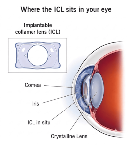
ICL surgery is an intraocular procedure that places a small, micro-thin prescription lens inside the eye, positioned behind the Iris (the coloured part) of the eye and in front of the natural lens. Unlike traditional contact lenses, an ICL remains inside the eye almost permanently (though it can be removed or replaced if necessary) and works in harmony with the natural lens to focus light accurately onto the retina.


Why ICL surgery matters

Refractive errors like myopia (short-sightedness) are a growing global and local concern:  

  • Childhood Prevalence: A systematic review of Malaysian studies reported childhood myopia at 17.18% overall.  
  • Adolescent Spike: Prevalence increases sharply to 42.71% among adolescents aged 13 to 18 years.1
  • Demographic Factors: Higher rates were observed among females and the Chinese population in the reviewed cohorts.  

For adults seeking freedom from heavy spectacle frames or contact lens discomfort, ICL offers a proven alternative—especially when laser vision correction (like LASIK, KLEx or PRK) is anatomically unsuited.


Causes and Risk Factors

ICL surgery does not treat an infection or disease. It corrects a light focusing issue from the ciliary sulcus part of the eye like how a contact lens does on the surface cornea of the eye. These are the common reasons a person may be assessed for ICL surgery:

  • The eyeball is too long. Light focuses in front of the retina instead of directly on it, so far objects look blurred; this is called myopia, or short-sightedness. 
  • The Cornea (front clear window) of the eye is unevenly curved. Light focuses unevenly, so vision may look blurred or distorted at more than one distance; this is called astigmatism. 
  • The spectacle power is high. Stronger prescriptions may make glasses thicker or contact lenses less comfortable, so lens-based correction may be discussed. 
  • The cornea may not be ideal for laser reshaping. Some laser procedures work by changing the cornea, the clear front layer of the eye; if the cornea is too thin or irregular, the specialist may recommend another option instead. 
  • There may be inherited or demographic risk factors for myopia. The Malaysian review reported higher myopia prevalence among females and among the Chinese population in the included studies, but individual suitability still depends on the patient’s own eye measurements.
  • The internal eye space and eye health must be safe for a lens. The U.S. Food and Drug Administration patient booklet for EVO ICL lists several situations where surgery should not be done, including pregnancy or nursing, significant glaucoma, and not meeting the required corneal cell safety level as determined by the doctor. 
  • Patient looking for an alternative to laser vision correction.


Classification: what type of vision problem is being corrected?

For patients, the most useful “classification” is not a disease stage, but the type of focusing problem and which treatment pathway fits safely.

1. Myopia, or short-sightedness

Near objects may look clear, while far objects look blurred. ICL surgery may help by adding a prescription lens inside the eye so light focuses more clearly on the retina. The U.S. Food and Drug Administration’s 2022 EVO ICL page states that the lens is placed behind the iris and in front of the natural lens, and does not replace the natural lens.

2. Myopia with astigmatism

Vision may be blurred or distorted because the eye focuses light unevenly. A toric ICL is a version designed to correct astigmatism as well as short-sightedness. The FDA’s 2022 approval page describes EVO/EVO+ Visian Toric ICL as intended for patients with near-sightedness and astigmatism.

3. Presbyopia, or age-related near focusing difficulty

Some patients in their 40s and above also notice difficulty reading up close. This is a separate focusing issue from myopia. ICL may not remove the need for reading glasses in every patient, and the best option depends on age, near-vision needs, cataract status, and eye measurements.

4. Cataract or early lens changes

If the natural lens is already cloudy, the discussion may shift away from ICL and toward cataract or lens-based surgery. This is because ICL keeps the natural lens in place, while cataract surgery removes and replaces the cloudy natural lens.

In the FDA clinical study summarised on its 2022 device page, 327 patients were followed; at six months, 75.9% of implanted eyes achieved 20/20 vision or better, and 98.9% achieved 20/32 vision.2  These results are encouraging, but they are not a guarantee for every patient. 

ConditionVisual ImpactHow ICL Helps
MyopiaNear objects are clear; far objects are blurry.Adds a corrective lens in front of the natural lens to focus light on the retina.
AstigmatismLight focuses unevenly, causing distorted vision.Toric ICL models align with the eye’s axis to neutralize astigmatic distortion.
PresbyopiaAge-related loss of close-up focusing (40+ years).Standard ICL does not stop presbyopia; reading glasses or monovision strategies or EDoF* ICL may be required.
CataractClouding of the eye’s natural lens.ICL is not suitable; treatment shifts to cataract surgery (lens replacement).


Signs and Symptoms

What can an Ophthalmology Clinician detect?

A patient cannot reliably check ICL suitability at home. During an assessment, the ophthalmology clinical team may check:

  • The exact prescription and whether it is stable. 
  • The corneal shape and thickness. 
  • The depth and shape of the front part of the eye. 
  • The health of the corneal endothelial cells, which help keep the cornea clear. 
  • Eye pressure. 
  • The retina, especially in patients with high myopia. 
  • Signs of cataract, glaucoma, inflammation, or other eye disease. 

A 2022 U.S. FDA clinical trial report on EVO ICL for moderate myopia measured vision, prescription accuracy, eye pressure, corneal endothelial cell density, and adverse events, showing why the assessment is more than just checking spectacle power.2 

What a patient with refractive error might notice at home/outdoor?

You may notice:

  • Blurred distance vision. 
  • Squinting to see far objects. 
  • Headaches or tired eyes. 
  • Glare or halos around lights. 
  • Difficulty driving at night. 
  • Contact lens induced dryness or intolerance. 
  • A frequent need to update glasses or contact lenses. 

The National Eye Institute lists blurry vision, glare or halos, squinting, headaches, eye strain, and focusing difficulty as symptoms of refractive errors. Seek an eye assessment if these symptoms appear, worsen, or remain troublesome despite glasses or contact lenses.

Urgent Red Flags: Patients must seek immediate care if experiencing a sudden burst of floaters, light flashes, or a dark shadow over vision, which may signal a retinal tear or detachment.  


Treatment Options

1. Glasses

What it is: Simple, non-invasive prescription lenses worn in a frame.
What it is for: Myopia, astigmatism, hyperopia, and presbyopia.
Limitations: High powers cause image distortion, weight, and limited peripheral fields.

2. Contact lenses

What it is: Prescription lenses worn on the surface of the eye.
What it is for: Patients who want a wider field of view or less dependence on glasses.
Limitations: Contact lenses require careful hygiene and regular replacement. Some patients develop dryness, irritation, allergy, or recurrent discomfort.

3. Laser vision correction

What it is: A laser reshapes the cornea, the clear front layer of the eye, so light focuses more accurately.
What it is for: Suitable for patients with appropriate corneal thickness, corneal shape, prescription range, tear film, and lifestyle needs.
Limitations: Not every cornea is suitable. Some patients may experience dry eye, glare, halos, under-correction, over-correction, or regression. Further re-treatment is sometimes considered, but “perfect” enhancement is not guaranteed.

4. ICL surgery

What it is: A small prescription lens is placed inside the eye, behind the iris and in front of the natural lens.
What it is for: Suitable for adults with myopia, with or without astigmatism, especially when laser correction is less suitable or when the prescription is high.
Limitations: ICL is an intraocular procedure, meaning surgery is performed inside the eye. Risks include glare, halos, raised eye pressure, cataract, corneal endothelial cell loss, inflammation, infection, retinal detachment, lens rotation, or need for lens removal or exchange. The FDA patient booklet specifically warns that cataract risk can rise with time after EVO ICL implantation and that the corneal endothelial cells need long-term monitoring. 

In a 2022 report of 200 eyes with moderate myopia from the U.S. FDA clinical trial, 91.5% were within ±0.50 dioptres of target and 100.0% were within ±1.00 dioptre at six months; mean endothelial cell density declined by 2.2%. These are study results, not a promise of the same result for every patient. 

5. Refractive lens exchange or cataract surgery

What it is: The natural lens is removed and replaced with an artificial lens implant.
What it is for: Patients with cataract, early lens changes, or selected older patients where lens-based correction is more appropriate than corneal laser or ICL.
Limitations: This is a different category of surgery from ICL because it removes the natural lens. It may change near focusing ability and carries its own surgical risks. Suitability depends on age, cataract status, retina health, prescription, and visual goals.


Screening and Risk Reduction

Before ICL surgery, screening is essential. The assessment usually includes prescription testing, eye pressure measurement, corneal scans, anterior-chamber measurements, endothelial cell assessment, crystalline lens inspection and a dilated retinal examination. The aim is not to “pass” or “fail” a simple number, but to decide whether the lens can be placed safely and whether another option may be better.


Post-Operative Care

Regular follow-up after ICL is also important. The lens may be designed for long-term placement, but the eye can change with time. Follow-up allows the doctor to monitor eye pressure, lens position, cataract development, corneal cell health, and retina health. The FDA patient booklet advises regular eye examinations after EVO ICL surgery to monitor cataract risk and corneal health.2 

Important patients’ note: ICL surgery does not stop myopia-related eye risks from existing. Patients with high myopia may still need periodic retinal checks even if their day-to-day vision improves.


Medical Disclaimer

This article is for general informational purposes only and is not a substitute for professional medical advice, diagnosis, or treatment. If you experience sudden double vision with headache, weakness, slurred speech, or facial drooping, seek emergency medical care immediately.


References and Further Reading

  1. Tan, E. S. Y., Agarwal, P., & Teng, C. L. (2026). Prevalence of myopia in children and adolescents: A systematic review of Malaysian prevalence studies. Malaysian Journal of Medical Sciences, 33(1), 14–26. https://doi.org/10.21315/mjms-08-2025-592

  2. U.S. Food and Drug Administration. (2022, April 18). EVO/EVO+ VISIAN Implantable Collamer Lens – P030016/S035.https://www.fda.gov/medical-devices/recently-approved-devices/evoevo-visian-implantable-collamer-lens-p030016s035

Laser Eye Surgery Side Effects: What to Ask During Your Consultation

Laser Eye Surgery Side Effects: What to Ask During Your Consultation

Laser Eye Surgery Side Effects: What to Ask During Your Consultation

Reviewed by Dr Diymitra K Ganasan, Refractive Surgeon of OasisEye Specialists | Last updated: July 2026


Laser eye surgery has helped millions of people see clearly without glasses or contact lenses — but like any procedure, it comes with possible side effects. The good news is that most are mild, temporary, and well understood. Your consultation is where you get to find out exactly how they apply to
your eyes, so you can go into surgery with realistic expectations rather than surprises.


What Are the Most Common Laser Eye Surgery Side Effects?

These are the side effects most patients are likely to experience, at least briefly, during the healing process:

  • Dry eyes — the most frequently reported side effect. Laser surgery can temporarily reduce tear production and corneal sensitivity, leading to a gritty or dry feeling that’s usually managed with lubricating eye drops while the eye heals.
  • Glare, halos, and starbursts — rings or streaks of light around bright sources, especially noticeable when driving at night in the first weeks after surgery.
  • Fluctuating or blurry vision — vision often improves day by day rather than instantly, with some blurriness as swelling settles and the cornea stabilizes.
  • Light sensitivity — temporary increased sensitivity to bright light or sunlight during the initial healing period.
  • Under-correction or over-correction — your vision may end up slightly less or more corrected than planned, but usually are able to provide functional vision. In very rare cases, a touch-up procedure (enhancement) may be required later.


Are Laser Eye Surgery Side Effects Permanent?

For most patients, no. FDA studies show a 95%+ satisfaction rate, with fewer than 1% reporting severe daily visual symptoms (U.S. Food and Drug Administration [FDA], 2021). Mild dry eye is the most common temporary issue, affecting a quarter of patients at three months before improving. Side effects are generally a normal, temporary part of healing—your specific risks depend on your eye profile, which is exactly what your consultation evaluates.


How Side Effects Compare Across Procedures

Not all laser vision correction is the same, and the side-effect profile varies by technique:

ProcedureKey Side-Effect Considerations
FemtoLASIKFast visual recovery; involves a corneal flap, so carries a small added risk of flap-related complications
SMILE pro 2.0Flapless and minimally invasive; generally associated with less dry eye than flap-based LASIK
TransPRKFlapless and bladeless; no flap risk at all, though surface healing takes a few days longer with more initial discomfort
ICL (Implantable Collamer Lens)Does not reshape the cornea at all, making it an option for thin corneas or very high prescriptions; carries its own distinct (and different) risk profile, such as lens-related concerns rather than corneal ones


Your surgeon will recommend a technique based on your cornea’s thickness and shape, your prescription, your lifestyle, and your tolerance for different recovery timelines — which is exactly why this should be a two-way conversation, not a one-size-fits-all recommendation.


What to Ask During Your Consultation

A good consultation is your chance to understand your personal risk profile, not just hear a general pitch. Here’s what’s worth asking.

About Your Candidacy

  • Am I a good candidate for laser eye surgery based on my corneal thickness and shape?
  • What do my corneal topography and pachymetry (corneal mapping and thickness) results show?
  • Do I have any dry eye, large pupils, or other factors that increase my personal risk of side effects?
  • Is my prescription stable, or could it still be changing?

About the Procedure Itself

  • Which procedure — FemtoLASIK, SMILE pro 2.0, TransPRK, or ICL — do you recommend for me, and why?
  • What technology will be used, and how does it reduce the risk of glare or halos?

About Risks and Side Effects

  • Based on my eyes specifically, which side effects am I most likely to experience?
  • How are dry eyes managed after surgery, and for how long might I need treatment?
  • What happens if I’m under-corrected or over-corrected — is an enhancement included, and what does it cost?
  • What are the signs that something isn’t healing normally, and who do I contact if that happens?

About Recovery and Aftercare

  • How long is the typical recovery period before I can drive, work, or exercise again?
  • How many follow-up visits are included, and over what timeframe?
  • Are there activities I should avoid, and for how long?

About Cost and Guarantees

  • Is the quoted price all-inclusive, or are follow-ups and potential enhancements billed separately?
  • What is your policy if I need an enhancement procedure later?


When to Seek Follow-Up Care After Surgery

Most healing follows a predictable pattern, but contact your surgeon promptly if you notice sudden pain, a marked drop in vision, increasing redness, or discharge at any point after surgery — these aren’t typical and deserve prompt review rather than waiting for your next scheduled visit.

Frequently Asked Questions About Laser Eye Surgery Side Effects

Dry eyes after laser eye surgery typically improve within three to six months as the cornea heals and nerve sensitivity is restored. Lubricating eye drops are usually prescribed throughout this period. Patients with pre-existing dry eye may take longer to recover, which is why a dedicated tear film assessment is part of the pre-operative screening process at OasisEye Specialists.

Recovery varies by procedure. Most FemtoLASIK and SMILE Pro 2.0 patients return to desk work within one to three days. TransPRK patients may need three to five days, as the corneal surface takes longer to heal. Roles involving dust, fumes, or prolonged screen time may require a longer rest period. Your surgeon will advise based on your specific procedure and work environment.

Post-operative drops typically include an antibiotic to prevent infection, an anti-inflammatory to support healing, and lubricating drops to manage dryness. Antibiotic and anti-inflammatory drops are usually continued for one to two weeks; lubricating drops may be recommended for several months depending on recovery. Your surgeon will prescribe a specific regimen tailored to your procedure and individual healing response.

Yes, laser eye surgery can correct astigmatism in suitable candidates. FemtoLASIK, SMILE Pro 2.0, and TransPRK can all treat myopic astigmatism within certain prescription and corneal parameters. The degree correctable depends on corneal thickness, shape, and the laser platform used. Suitability is confirmed through corneal topography, pachymetry, and a full refractive assessment during your consultation.

SMILE Pro 2.0 and TransPRK are both flapless but treat the cornea differently. SMILE Pro 2.0 extracts a small internal lenticule through a tiny incision and is generally associated with less post-operative dry eye and faster visual stabilisation. TransPRK treats the corneal surface directly, resulting in a slightly longer surface-healing period with more initial discomfort in the first few days. Individual suitability depends on corneal measurements and prescription profile.

Flap-related complications are uncommon with modern FemtoLASIK but remain a known consideration. These include flap dislocation from eye trauma, epithelial ingrowth along the flap edge, and diffuse lamellar keratitis — a post-operative inflammatory response. Rates are low with experienced surgeons and advanced laser platforms. For patients where flap-related risk is a concern, flapless alternatives such as SMILE Pro 2.0 or TransPRK may be more appropriate.


Ready to Discuss Your Options?

Laser eye surgery is elective, which means you have the time to ask every question that matters to you before deciding. A thorough consultation — including corneal mapping, a detailed discussion of your personal risk factors, and an honest comparison of which procedure suits your eyes — is the best way to walk into surgery confident and well-informed.


Medical Disclaimer

This article is for general informational purposes only and is not a substitute for a personalised consultation with a qualified ophthalmologist. Individual risk factors vary, and only a comprehensive eye examination can determine your suitability for laser eye surgery.

What Age is Best for Laser Eye Surgery and How It Affects The Types of Treatment Decision by Doctor?

Asian mother and daughter wearing glasses, smiling together indoors, representing different age groups for laser eye surgery considerations

General Age Guidelines

Close-up of human brown eye with clear cornea and lashes.

  1. Minimum age: 21 years old
  2. Stability of prescription: Doctors want a stable refraction (no major changes in glasses power).
  3. No other corneal anomalies, like past history of cornea scar or injury or signs of corneal diseases
  4. Upper age limit: There isn’t a strict cutoff, but past 40–45 years, presbyopia (age-related near blur) and early lens changes start influencing treatment choice.


Treatment Options by Age Group

Young Asian man with glasses smiling and holding coffee.

21-40 years 

The cornea is generally still healthy, healing capacity is good.

If the prescription is stable:

  1. TransPRK, FemtoLASIK, SMILE Pro: preferred for low-to-moderate myopia and astigmatism.
  2. ICL considered if: very high myopia, thin cornea, or unsuitable corneal shape.

Doctors may delay surgery if frequent prescription changes are noted.

Woman holding eyeglasses with blurred face in background.

40-50 years (Onset of Presbyopia)

The natural lens stiffens, need for reading glasses.

Laser options (SMILE pro, FemtoLASIK, TransPRK) still possible, but patients must be counseled about:

  • Reading glasses after surgery
  • Or monovision (one eye corrected for distance, one for near)
  • Another option, PRESBYOND, this treatment uses special technology to extend the range of eye natural focus range (Depth of focus) to allow reading at normal reading distance 
  • ICL: still possible, especially for high myopia.

Presbyopic IPCL: suitable for presbyopia patients.

Some patients may start showing early cataract signs, refractive lens exchange (RLE) might be discussed instead.

Elderly Asian man adjusting glasses while looking at smartphone.

Above 50 years

Higher likelihood of cataracts developing.

Doctors may recommend lens-based surgery (RLE or cataract surgery with premium lens implants) instead of corneal laser procedures.

ICL is less favored after 50, since cataract surgery may be needed soon anyway.

Most patients in this age range benefit more from lens replacement than laser on the cornea.

See a refractive surgeon and get a detailed evaluation to see what can realistically be achieved. The refractive surgeons available in OasisEye Specialists include: 

In between SMILE pro, FemtoLASIK, TransPRK – Which one is for you?

Choosing the right laser eye surgery can feel overwhelming, especially with options like SMILE pro, TransPRK, and FemtoLASIK. Each procedure is designed to correct vision and reduce or eliminate the need for glasses and contacts, but the best choice depends on your eye condition, lifestyle, and vision goals. Here’s a breakdown of each option to help you decide.

What is SMILE pro?

SMILE pro (Small Incision Lenticule Extraction) is a minimally invasive procedure that corrects myopia and astigmatism. Unlike LASIK, it doesn’t involve creating a corneal flap. Instead, a small incision is made in the cornea to remove a lenticule, reshaping the cornea and improving vision.

  • Best for: Individuals with myopia and mild astigmatism seeking a minimally invasive procedure with fast recovery.
  • Benefits: Quick recovery, less dry eye risk, no flap-related complications.
  • Considerations: Limited to certain prescriptions (myopia and mild astigmatism).

What is TransPRK?

TransPRK (Transepithelial Photorefractive Keratectomy) is a surface-based laser surgery that does not require an incision or flap. Instead the laser simply ablates (surgically removes) the outermost layer of cells on the surface of the eye to reach the cornea. This makes it an excellent choice for individuals with thin corneas or those who aren’t eligible for FemtoLASIK or SMILE pro. TransPRK offers more versatility by treating a wider range of refractive errors, including myopia, hyperopia, and astigmatism. However, it comes with a longer recovery time and more post-surgery discomfort, as the outer layer of the cornea takes time to regenerate.

  • Best for: Patients with thin corneas, irregular corneas, or those wanting to avoid any cuts or flaps.
  • Benefits: No flap creation, suitable for thin corneas, less invasive.
  • Considerations: Longer recovery time and more discomfort during healing.

What is FemtoLASIK?

FemtoLASIK is a popular procedure where a femtosecond laser creates a corneal flap. After the flap is lifted, an excimer laser reshapes the cornea to correct the vision. The flap is then repositioned, and recovery begins.

  • Best for: Those seeking fast recovery, minimal discomfort, and who have suitable corneal thickness for flap creation.
  • Benefits: Quick visual recovery, minimal post-op discomfort.
  • Considerations: Slight risk of flap-related complications, such as displacement.

Which One Is Best for You?

The best choice between SMILE pro, TransPRK, and FemtoLASIK depends on several factors, including your eye health, lifestyle, and personal preferences.

  • SMILE pro is ideal for those with myopia and mild astigmatism who want a minimally invasive option with a fast recovery.
  • TransPRK is better suited for individuals with thin or irregular corneas or those who prefer a surface-based procedure without incisions or flaps.
  • FemtoLASIK offers the quickest recovery and is a great option for those with appropriate corneal thickness and who want minimal post-surgery discomfort.

Conclusion

Ultimately, the decision between SMILE pro, TransPRK, and FemtoLASIK depends on your unique eye conditions and vision goals. Consulting with an experienced ophthalmologist will provide personalized guidance on which procedure is the best fit for you.