
Reviewed by Dr Wilson Wong Jun Jie, Vitreoretinal and Refractive Surgeon of OasisEye Specialists | Last updated: October 2026
Myopic traction maculopathy is a vision-threatening structural disorder of severe short-sightedness in which progressive elongation of the eyeball stretches and splits the delicate layers of the central retina.
Understanding the Risks of Severe Short-Sightedness
Severe short-sightedness (high myopia) is far more than a requirement for strong prescription glasses; it represents a structural stretching of the entire eye. Global epidemiological models published in Ophthalmology (Holden et al., 2016) project that high myopia will affect nearly 1 billion people worldwide by 2050.
Malaysian studies have reported high-myopia prevalence ranging from 0.40% to 4.88% among children and adolescents, although differences in study populations and definitions prevent a reliable pooled national estimate (Tan et al., 2026). As the eye stretches significantly longer than normal, the thin wall at the back of the eye bulges outward, putting extreme physical tension on the light-sensitive nerve layer (the retina). Left unmonitored, this mechanical stretching can cause progressive internal tearing of the retina, leading to irreversible loss of detailed central vision.
Causes and Risk Factors
- Physical lengthening of the eye (Axial Elongation): As the front-to-back length of the eye grows abnormally long, the rigid outer wall stretches thin, creating an outward bulge at the back of the eye that pulls the retina apart.
- Outward bulging of the back eye wall (Posterior Staphyloma): An abnormal contour develops at the back of the eyeball, causing the curved outer wall to recede away from the flat, inelastic retina lining it.
- Inward pulling of internal eye gel (Vitreomacular Traction): The clear gel filling the eye shrinks and sticks to the central retina, creating inward pulling forces that pull against the outward stretching of the back eye wall.
- Stiffening of internal retinal membranes (Epiretinal Membrane Tension): Fine scar-like tissue forms on the surface of the retina, creating tangential surface tension that prevents the retina from resting comfortably against the wall of the eye.
Staging of Structural Retinal Stretching (Myopic Traction Maculopathy)
Clinicians categorize retinal stretching using the standardized Myopic Traction Maculopathy Staging System (MSS) (Parolini et al., 2021). The practical implications for each stage include:
- Stage 1 (Inner or Outer Layer Splitting / Maculoschisis): The layers inside the retina have begun to separate fine microscopic layers, but the primary visual cells remain attached. Vision may feel normal or show mild distortion.
- Stage 2 (Pure Outer Layer Splitting / Foveoschisis): The splitting has spread into the deepest layers of the central vision zone. Patients often notice distortion or difficulty reading fine print. This stage represents the primary surgical decision threshold for structural intervention.
- Stage 3 (Central Retinal Detachment / Foveal Detachment): The light-sensing cells have pulled entirely away from the back wall of the eye in the central visual area. Central vision drops noticeably, and prompt surgical intervention is required to prevent cell death.
- Stage 4 (Full Posterior Retinal Detachment): The fluid separation extends across the entire back pole of the eye. Central vision is significantly impaired, and complex restorative surgery is necessary to preserve basic functional sight.
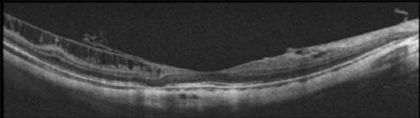
Image A: Stage 1 - Maculoschisis
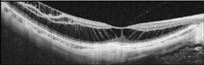
Image B: Stage 2 - Foveoschisis
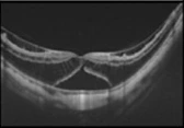
Image C: Stage 3 – Foveal Detachment
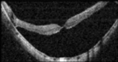
Image D: Stage 4 – Full Posterior Retinal Detachment

Signs and Symptoms
Because early retinal splitting occurs inside the microscopic layers of the eye, early stages are frequently invisible without specialized clinic imaging.
What Only a Clinician Can Detect During Screening
- Microscopic separation between internal retinal layers on high-resolution Optical Coherence Tomography (OCT) cross-sectional scans.
- Subtle fluid pockets under the central retina (subretinal fluid).
- Early structural formation of a posterior staphyloma (outward bulging of the sclera).
- Microscopic full-thickness or partial-thickness holes in the central macula (lamellar or full-thickness macular holes).
What a Patient or Family Member Might Notice at Home
- Straight lines (such as door frames, window blinds, or lines of text) appearing bent, wavy, or kinked.
- A dark, hazy, or blurred spot in the exact center of vision that does not clear with blinking or changing reading glasses.
- Difficulty reading small print or recognizing faces, even when wearing updated glasses.
- Sudden appearance of persistent dark spots (floaters) or flashes of light.
Important Safety Instruction: If you or a family member notice lines appearing wavy or a persistent dark blur in your central vision, seek an immediate clinical evaluation by a retinal specialist. Delayed evaluation and intervention are associated with a higher risk of permanent visual loss.
Treatment Options for Severe Myopic Retinal Stretching
1. Non-Penetrating Macular Buckle (NPB Surgery)
- What It Is: An advanced surgical procedure where a tiny, custom-shaped biocompatible implant (made of medical-grade PMMA coated in silicone) is placed gently against the outside back wall of the eyeball.
- What It Is For: It supports and pushes the bulged outer wall of the eye inward, neutralizing the physical stretching forces pulling the retina apart.
- Limitations & Risks: Visual recovery is gradual, taking between 1 to 6 months as fluid slowly absorbs. While anatomical reattachment rates exceed 80–90% in published series (Parolini et al., 2023 meta-analysis), long-term visual improvement depends on the health of the visual cells prior to surgery. Advanced pre-operative damage limits ultimate visual potential.
2. Pars Plana Vitrectomy (PPV Surgery)
- What It Is: A micro-incisional internal surgery where the vitreous gel inside the eye is carefully removed and microscopic membranes pulling on the front surface of the retina are peeled away.
- What It Is For: Relieves inward pulling forces coming from inside the vitreous cavity.
- Limitations & Risks: In eyes with severe outward bulging (posterior staphyloma), internal gel removal alone may not resolve the physical stretching coming from behind the eye.
3. Combined Macular Buckling and Vitrectomy
- What It Is: A dual-approach procedure combining external buckle support with internal gel removal and membrane peeling.
- What It Is For: Used in advanced Stage 3 or 4 cases, or when a full-thickness macular hole accompanies severe retinal detachment.
- Limitations & Risks: Requires longer operating time and careful post-operative monitoring. Gas or oil bubbles placed inside the eye during surgery may require temporary face-down positioning.
Screening and Prevention Guidelines
While structural eye lengthening cannot be reversed once it occurs, population-level evidence shows that the majority of severe vision loss cases are associated with delayed screening and late treatment.
- Annual High-Resolution Retinal Scans: Individuals with high myopia (prescription greater than -6.00 Dioptres or axial length over 26 mm) should undergo annual Optical Coherence Tomography (OCT) scanning to check the deep retinal layers.
- Home Amsler Grid Monitoring: High myopes should check each eye individually once a week using an Amsler grid (a grid of straight lines) to catch early line distortion at home.
- Baseline Retinal Mapping: Young adults with high myopia should establish a baseline structural map of their eye wall to track subtle changes over time.
Medical Disclaimer
This article is for general informational purposes only and is not a substitute for a personalised consultation with a qualified ophthalmologist. Individual risk factors vary, and only a comprehensive eye examination can determine your suitability for laser eye surgery.
References
Holden, B. A., Fricke, T. R., Wilson, D. A., Jong, M., Naidoo, K. S., Sankaridurg, P., Wong, T. Y., Naduvilath, T. J., & Resnikoff, S. (2016). Global prevalence of myopia and high myopia and temporal trends from 2000 through 2050. Ophthalmology, 123(5), 1036–1042. https://doi.org/10.1016/j.ophtha.2016.01.006
Parolini, B., Arevalo, J. F., Hassan, T., Kaiser, P., Rezaei, K. A., Singh, R., Sakamoto, T., Rocha, J., & Frisina, R. (2023). International validation of myopic traction maculopathy staging system. Ophthalmic Surgery, Lasers and Imaging Retina, 54(3), 153–157. https://doi.org/10.3928/23258160-20230217-01
Parolini, B., Palmieri, M., Finzi, A., Besozzi, G., Lucente, A., Nava, U., Pinackatt, S., Adelman, R., & Frisina, R. (2021). The new myopic traction maculopathy staging system. European Journal of Ophthalmology, 31(3), 1299–1312. https://doi.org/10.1177/1120672120930590
Tan, E. S. Y., Agarwal, P., & Teng, C. L. (2026). Prevalence of myopia in children and adolescents: A systematic review of Malaysian prevalence studies. Malaysian Journal of Medical Sciences, 33(1), 14–26. https://doi.org/10.21315/mjms-08-2025-592
Frequently Asked Questions (FAQ)
A traditional scleral buckle is a silicone band placed around the equator (middle portion) of the eye to treat peripheral retinal tears. A macular buckle (such as the NPB device) is specifically engineered with a specialized curved arm to reach the very back center of the eyeball (the macula) to support outward bulging caused by high myopia.
The surgery is performed under localised or general anesthesia, so you will not feel pain during the procedure. Post-operative discomfort is typically mild to moderate and managed with prescribed eye drops and oral analgesics.
Initial surgical healing occurs over 2 to 4 weeks. However, structural visual recovery is a gradual process that typically takes between 1 and 6 months as the delicate retinal layers slowly re-settle onto the supported eye wall.
Laser surface procedures like LASIK, SMILE Pro 2.0, or TransPRK reshape the front surface (cornea) of the eye to reduce your glass prescription. However, they do not change the physical length of the eyeball or treat structural stretching at the back of the eye (retina). A comprehensive retinal evaluation is mandatory before considering any refractive surgery.
The primary goal of macular buckling is to stabilize the structure of the eye and prevent progressive, irreversible vision loss. While most patients experience stabilization or improvement in distortion, final visual acuity depends on how long the central retina was stretched or detached prior to surgery.