Reviewed by Dr Khaw Hoon Hoon, Refractive Surgeon of OasisEye Specialists | Last updated: October 2026
Understanding Refractive Errors: Context & Impact
Refractive errors—primarily near-sightedness (myopia), far-sightedness (hyperopia), and irregular corneal curvature (astigmatism)—affect a vast segment of the global population. According to World Health estimates published in Ophthalmology (Holden et al., 2016), approximately 30% of the global population currently has myopia, a figure projected to rise significantly in urbanized regions.
In Malaysia and across East and Southeast Asia, the prevalence of myopia among young adults in urban centers is particularly high, often exceeding 60% to 80% (Dong et al., Journal of Ophthalmology, 2020). For many individuals, relying on thick glasses or contact lenses introduces daily inconvenience, dry eye discomfort, or lifestyle restrictions in sports and outdoor activities. Laser vision correction offers a permanent, surgical option to reshape the clear front window of the eye (the cornea) so that light rays focus accurately onto the retina.
Causes and Risk Factors
Refractive errors develop when there is a mismatch between the bending power of the eye’s optical components and the physical length of the eyeball.
- Elongated Eyeball Structure (Axial Myopia): The physical front-to-back length of the eye grows longer than average, causing light rays to focus in front of the retina rather than directly on it.
- Irregular Corneal Curvature (Astigmatism): The clear front dome of the eye is shaped more like an oval football than a round basketball, causing light to focus at multiple different points inside the eye.
- Genetic Predisposition: Having one or both parents with high nearsightedness or astigmatism is strongly associated with an increased likelihood of developing similar focus issues (Tideman et al., JAMA Ophthalmology, 2018).
- Environmental and Lifestyle Factors: Prolonged near-work activity and limited time spent outdoors during childhood development are linked to higher rates of myopia progression (Morgan et al., Progress in Retinal and Eye Research, 2012).
Severity & Staging: What Refractive Error Means for You
In clinical practice, refractive errors are measured in diopters (D)—the unit used to express the optical power of a lens. Understanding your level of refractive error helps determine your eligibility for specific types of vision correction procedures.
- Mild Refractive Error Up to -3.00D): Uncorrected vision is slightly blurry at a distance. Daily tasks without glasses are often manageable, though driving or reading signs requires correction.
- Moderate Refractive Error (-3.00D to -6.00D): Distance vision is significantly impaired without glasses or contact lenses. Facial features or objects beyond arm’s length appear blurred.
- High Refractive Error (Greater than -6.00D): Clear vision is limited to objects positioned just a few inches from the face. High myopia is also associated with structural changes inside the eye that require routine retinal monitoring.
Signs and Symptoms
Identifying a change in your eyesight involves recognizing how your vision performs in daily settings compared to what a doctor evaluates during a comprehensive exam.
What You Might Notice at Home
- Blurry vision when looking at distant objects, road signs, or presentation screens.
- Squinting or screwing up the eyes to see clearly.
- Eye strain, fatigue, or dull headaches after driving or reading.
- Distorted, double, or shadowed letters when reading text (often indicative of astigmatism).
- Difficulty seeing clearly in low-light or nighttime conditions.
What Only a Clinician Can Detect During an Exam
- Exact optical prescription and astigmatic axis measured via digital autorefraction and subjective testing.
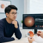
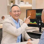
- Corneal thickness and 3D structural mapping (corneal topography/tomography) to ensure structural stability.
- Dry eye parameters and tear film stability.
- Microscopic evaluation of the crystalline lens and detailed peripheral retina examination.
Treatment Options: The Evolution of Laser Vision Correction
Laser vision correction has evolved significantly over the last three decades, moving toward increasingly minimally invasive techniques.
| Procedure | PRK (Photorefractive Keratectomy) | LASIK (Laser-Assisted In Situ Keratomileusis) | First-Generation SMILE (Small Incision Lenticule Extraction) | ZEISS SMILE pro 2.0 (At OasisEye Specialists) |
|---|---|---|---|---|
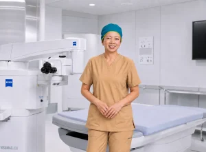
| Technique | Surface treatment; gentle removal of corneal epithelium, followed by excimer laser reshaping of exposed surface tissue. | Dual-laser flap procedure; femtosecond laser creates a hinged circular corneal flap, lifted for excimer laser reshaping before replacement. | First-generation flapless keyhole procedure; ZEISS VisuMax 500 femtosecond laser forms a tissue lenticule removed through a 2 mm incision. | Advanced AI-assisted flapless procedure using the ZEISS VisuMax 800 platform with automated alignment and topography integration. |
| Primary Indications | Thin corneas, superficial corneal scars, or structural surface irregularities unsuitable for flap/keyhole procedures. | Mild-to-high myopia, hyperopia, and astigmatism seeking rapid visual recovery. | Myopia and astigmatism; ideal for active lifestyles and dry-eye-prone eyes. | Precision correction of myopia and astigmatism with maximum patient comfort and ultra-fast treatment speed. |
| Laser Suction Time | N/A (Surface Excimer Laser) | ~15–20 seconds per eye for flap creation | ~25–30 seconds per eye | 7–10 seconds per eye |
| Key Advantages | Flapless; preserves maximal corneal thickness; avoids flap-related complications. | Rapid visual recovery within 24 hours; minimal post-operative discomfort. | Flapless; preserves anterior corneal biomechanics; significantly lower risk of post-operative dry eye. | Ultra-Fast: 7–10s laser phase reduces fixation risk. OcuLign: Compensates for eye rotation to maximize astigmatism accuracy. AI Planning: Customizes lenticule geometry. |
Limitations & Considerations for SMILE pro 2.0: As with all laser vision correction procedures, SMILE pro 2.0 does not prevent age-related reading vision changes (presbyopia) that occur naturally later in life. While regression (a slight return of prescription over time) is uncommon, studies report long-term regression rates of approximately 1% to 3% over 5 to 10 years depending on the initial prescription (Reinstein et al., Journal of Refractive Surgery, 2023).
Screening and Pre-Operative Assessment
Laser vision correction is an elective procedure, and thorough pre-operative screening is the most critical step in achieving a safe outcome.
- Comprehensive Corneal Mapping: Advanced 3D tomography measures corneal thickness, elevation, and structural symmetry to screen out pre-existing conditions such as keratoconus (a progressive thinning of the cornea).
- Prescription Stability: Candidates should generally demonstrate a stable optical prescription for at least 12 months prior to surgery.
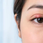
- Dry Eye & Ocular Surface Health: Pre-existing severe dry eye or eyelid inflammation must be managed prior to undergoing any laser procedure.
- Individualized Evaluation: Your ophthalmic specialist will determine eligibility based on current clinical guidelines set by international bodies, such as the American Academy of Ophthalmology (AAO Preferred Practice Pattern: Refractive Surgery, 2023), and local clinical protocols.