Hypertensive Retinopathy

The retina is the clear photosensitive tissue that is located at the back of the eyes. It helps to process and transform light into nerve signals. The nerve signals will be interpreted in the brain, providing visions. Serious medical conditions could affect the retina, such as high blood pressure (hypertension).

Hypertensive retinopathy (HR) is an eye disease in the retina due to complications of high blood pressure (hypertension). Poor control of high blood pressure may cause vascular disease changes without being aware of it. As a result, the retina’s blood vessel walls will thicken, causing the blood vessels to become narrow and restrict the blood flow reaching the retina. Over time, high blood pressure can limit the functioning of the retina by putting pressure on the optic nerve. Eventually, it can cause vision problems. For this reason, it is essential for patients with high blood pressure to have a regular eye check.

Older people are at a higher risk of developing high blood pressure and, therefore, most likely to develop hypertensive retinopathy. The following conditions can also increase your risk of hypertensive retinopathy: They include prolonged high blood pressure, heart diseases, atherosclerosis, diabetes, smoking, high cholesterol, obesity, having an unhealthy diet, having a family history of high blood pressure, and heavy alcohol consumption.
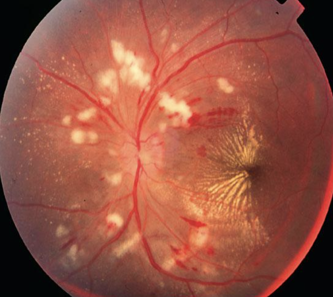
Fundus image of hypertensive retinopathy showing retinal hemorrhages, cotton wool spots, and optic disc swelling.

Symptoms and signs

Generally, a person with mild hypertensive retinopathy might not experience any or very mild symptoms. The patient usually becomes more symptomatic as it progresses and in the late stages. However, an ophthalmologist can detect early stages by identifying vascular changes during fundus examination.

  • Common symptoms include:
  • Blurred vision
  • Subconjunctival haemorrhage (bleeding in the white part of the eye)
  • Double vision accompanied by a headache

Controlling high blood pressure is the only way to treat hypertensive retinopathy. Therefore, lifestyle changes that include quitting smoking, exercising regularly, losing weight, dietary changes, and reducing alcohol intake help to reduce the risk of this disease.

However, individuals with hypertensive retinopathy are at risk of developing various complications. This includes the following:-

  • Retinal artery occlusion: It occurs when blood clots block the arteries that carry blood to the retina. This results in insufficient oxygen or blood supply in the retina, which may cause loss of vision.
  • Retinal vein occlusion: This condition occurs when a blockage in the retinal veins prevents blood from being carried away from the retina. A blood clot in the veins causes it.
  • Ischemic optic neuropathy: This condition occurs when high blood pressure blocks normal blood flow within the eyes and leads to damage to the optic nerve, which responsibly transmits images to the brain.
  • Malignant hypertension: This condition occurs when a sudden increase in blood pressure interferes with vision, causing sudden vision loss. However, this condition rarely happens but it could be potentially life-threatening.

Hypertensive retinopathy is also associated with an increased risk of stroke and heart attack. Prevention of hypertensive retinopathy is possible by carefully managing high blood pressure and related conditions like diabetes. Lifestyle changes such as quitting smoking, losing weight, exercising regularly, and having a healthy diet could also help prevent hypertensive retinopathy. People with high blood pressure should monitor their blood pressure regularly and have an annual eye screening. It is urged to visit an ophthalmologist as soon as possible if there is a significant sign and symptom of hypertensive retinopathy.
Macular Hole

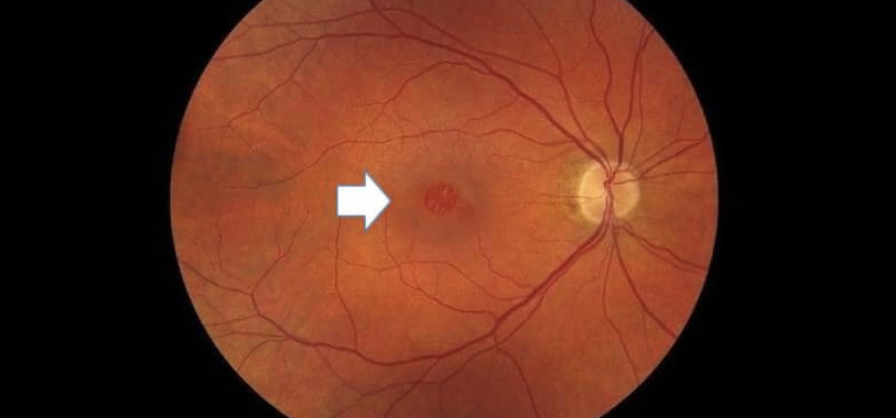
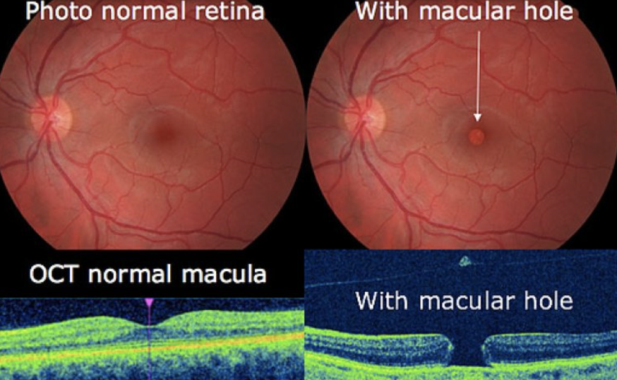
Macular hole occurs when there is a tear or hole that develops in the central part of your vision known as the macula. Macula is the small area of the retina where light is sharply focused which allows you to see colour and detailed vision. The common symptoms of a macular hole are distortion of vision (straight lines appear wavy), blurred vision or a dark spot in your central vision. You may not notice these symptoms with both eyes open. You should close one eye (better eye) and then you will notice straight lines appearing wavy and also a dark spot in the center of your vision. It is important to schedule an appointment with an Ophthalmologist as soon as possible if any of these symptoms occur. A special imaging test known as optical coherence tomography (OCT) which gives a cross sectional view of the retina will be performed by your eye doctor to diagnose the macular hole.

Fundus image of an eye with a macular hole, marked by a white arrow, showing central retinal damage.

Age is the most common cause for macular hole. As you get older, the vitreous starts to shrink and pull away from the retina. Usually the vitreous pulls away without causing any problems. Sometimes, the vitreous can stick to the retina and this causes the macula to stretch and a hole to form.

Comparison of normal retina and retina with a macular hole using fundus photos and OCT scans.

Treatment for macular hole

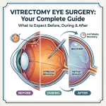
The treatment for a macula hole is by a surgery known as vitrectomy. Vitrectomy surgery is done by making three incisions into the sclera. The first incision is to make a hole for the infusion cannula where fluid is injected into the eye to keep the eyeball in shape. The second incision is for the light pipe to allow surgeons to see inside the eye. The last incision is for the vitrectomy cutter where it functions to remove and suck out the vitreous jelly from the eye. Vitrectomy is done by removing the vitreous jelly inside the eye that is pulling on your macula and filling the eye with a gas bubble. The bubble helps to flatten the macula hole and hold it in place until the eye heals. You should not fly or go uphill with a gas bubble in the eye until the gas dissipates on its own and is replaced by natural eye fluids. You will need to maintain a face-down position for several days to keep the gas bubble in place and help close the hole.

The complications of vitrectomy surgery are rare. Some complications include infections, bleeding, retinal detachment or recurrence of a macula hole. These complications can be easily treated. This operation is successful about 90-95% to close the hole. However, as the retina is a nerve tissue, it can take quite a few months to heal completely. People who have had a macular hole in one eye have about 10-15 percent chance of developing a macular hole in another eye over a lifetime. If you have symptoms such as blurred or distorted vision, do consult your eye care professional immediately. A relatively early treatment may give a better outcome in terms of improvement in your vision.

Floaters and Flashes

What are floaters?

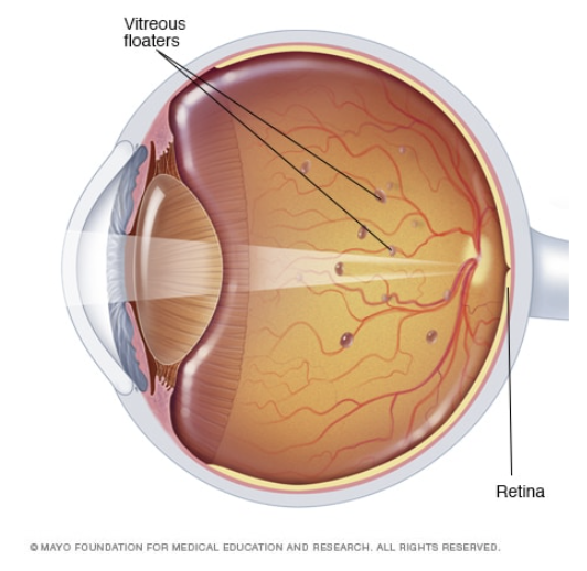
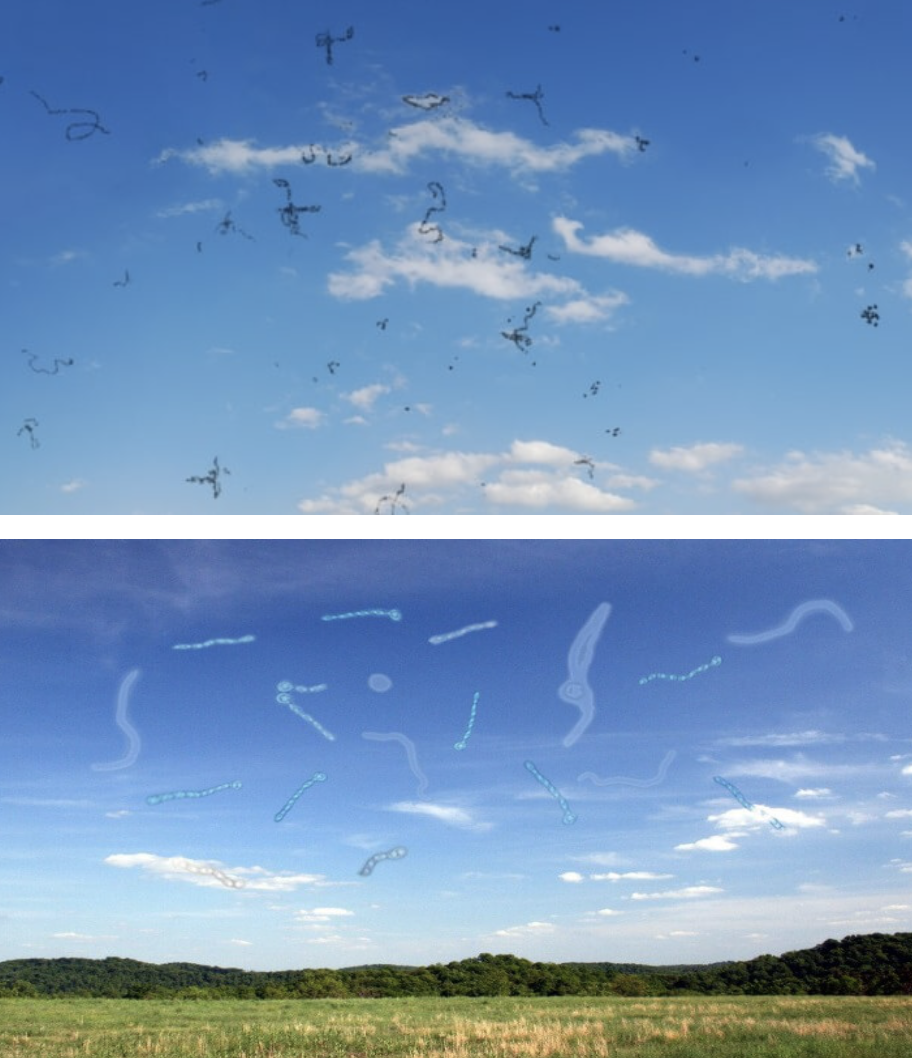
Have you ever noticed little tiny black dots in your vision? These are usually called floaters in layman’s terms. Floaters are usually more noticeable in a bright environment or a bright blue sky on a sunny day. Floaters can come in different shapes, such as little dots, circles, lines, clouds or cobwebs. It may be of a sudden onset and it is usually due to the vitreous which is the jelly inside your eye, shrinking due to age. This process occurs especially when you’re growing older or if you’re very short-sighted (myopia), then you would get it earlier. However, it can come about in other conditions as well, such as in diabetes and when you have an infection or inflammation in your eye. In most people, this is an innocuous or relatively minor problem that happens as you get older.

Eye diagram showing vitreous floaters and retina.

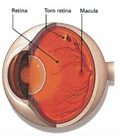
Floaters are normal and a common part of aging. However, at least 1 percent of the time, the shrinking of the vitreous or the jelly may happen. It starts with peeling off your retina. The retina is a thin layer inside your eye that is full of nerve tissues which is like the film in a camera and is what you use to see things. Now if this jelly shrinks and pulls on your retina too hard, it can sometimes cause a tear. If there is a tear, you will need treatment for it. The incidence of this is about 1 percent so most of the time nothing needs to be done. However, we also need to rule out other problems such as infection or inflammation or perhaps bleeding in the eye. While floaters are typically harmless, if it appears suddenly and are accompanied by flashes of light or impaired vision, it can signal a potentially serious eye problem.

Eye illustration highlighting a torn retina.

What are flashes of light?

Flashes look like flashing lights or lightning streaks in your field of vision. Flashes happen when the vitreous pulls on the retina. In younger people, the gel inside the eye (vitreous) is firmly attached to the retina. As you get older, the vitreous gel becomes more liquid and slowly collapses away from the retina. Flashes can be a symptom of a retinal tear or retinal detachment. A retinal tear is a break in the retina, while a retinal detachment is when the vitreous pulls away, creating a break and fluid from the vitreous gets behind the retina, causing blurred vision. These are serious conditions that can damage your sight. Flashes are usually seen in people as they age or in those with migraines. Flashes can appear on and off for several weeks or months. Flashes that are caused by migraine usually happen among younger people. When you have a migraine, it causes flashes that look like jagged lines or heat waves. This can occur in one or both eyes and can last up to 20 minutes.

Treatment

Treatment of floaters and flashes depends on the underlying condition. A thorough eye examination by an Ophthalmologist will determine if there are any damages to the retina. While some floaters will remain in your vision, many of them will fade over time and cause fewer problems. However, for large floaters that obstruct daily tasks, a vitrectomy surgery can be performed to remove the vitreous gel inside the eye and it will be replaced with a bubble of air or gas. The gas will disappear over time and will be replaced with a clear fluid produced by the eye.

Simulated vision of floaters in the sky..

What can you do about floaters and flashes in your eye?

It is important to take care of your eyes as you age. If you notice occasional eye floaters or flashes in your vision is not something you should worry about. This is normal and often happens when you age. An increased number of floaters in your vision or flashes should be a call for an emergency. When this occurs, it is wise to see an Ophthalmologist immediately to avoid any vision loss. Make an appointment to see an Ophthalmologist if you experience any of the warning signs below:

  • A new onset or increase in floaters or flashes of light
  • A sudden increase in size and number of floaters
  • A sudden change or decline in vision

Degenerasi Makula Berkaitan Usia (ARMD)

Degenerasi makula adalah penyakit yang berkait rapat dengan usia. Penyakit ini disebabkan oleh kerosakan pada bahagian makula.

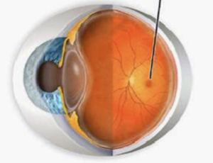
Diagram of an eye highlighting the macula, linked to AMD.Makula adalah satu kawasan kecil di bahagian tengah retina yang memiliki banyak sel penglihatan. Punca utama degenerasi makula kering masih tidak dapat dikenalpasti. Tetapi penyelidikan menunjukkan ianya mungkin dipengaruhi oleh faktor genetik dan factor persekitaran, termasuk merokok, obesiti dan diet. Apabila umur semakin meningkat, bermula pada usia 55 tahun dan keatas sesetengah berlakunya perubahan negatif pada makula. Kekurangan zat juga menyebabkan sel-sel di makula rosak dan tidak dapat berfungsi dengan baik. Perubahan ini menyebabkan penglihatan menjadi kabur sehingga menjadi buta.

Jenis-jenis Degenerasi Makula

  1. Degenerasi Makula Kering
    Lapisan makula menipis dan terbentuk mendapan lemak berwarna kuning yang dikenali sebagai “Drusen’. Kebiasaannya pada peringkat ini penglihatan akan terjejas apabila bilangan “drusen” semakin banyak dan saiz semakin besar.
  2. Degenerasi Makula Basah
    Salur darah tidak normal akan terbentuk di makula. Ini akan menyebabkan pendarahan berlaku dan penglihatan menjadi kabur. Meskipun jenis ini boleh mengakibatkan hilang penglihatan yang teruk, namun ianya jarang menyebabkan buta sepenuhnya.

Bagaimana pesakit degenerasi makula melihat?

Tanda-tanda awal yang mungkin anda perhatikan adalah kualiti penglihatan berkurang secara beransur-ansur atau garis lurus kelihatan bengkok dan senget. Apabila degenerasi makula berkembang, penglihatan pusat akan menjadi kabur. Oleh itu mereka mengalami kesukaran untuk membaca, mengenali wajah dan memandu. Mereka juga sensitif pada cahaya yang terang. Namun begitu mereka yang mengalami penyakit ini tidak akan mengalami kebutaan keseluruhan kerana penglihatan sisi (periferi) masih kekal.

Bolehkah degenerasi makula disembuhkan?

Walaupun tidak ada penawar untuk AMD, terdapat pilihan rawatan yang dapat mencegah atau melambatkan perkembangan penyakit ini. Rawatannya merangkumi ubat-ubatan, suntikan dan terapi laser. Rawatan terbaik untuk makula degenerasi basah adalah terapi anti-VEGF- iaitu suntikan bahan kimia yang disebut “anti-VEGF” secara intravitreal (ke dalam mata). Dalam kehidupan normal tubuh manusia, VEGF adalah molekul sihat yang menyokong pertumbuhan darah baru. Rawatan ini dapat membantu menghentikan kebocoran salur darah yang disebabkan oleh makula degenerasi basah.

Bagaimana anda mencegah degenerasi makula menjadi lebih teruk?

  • Mengekalkan berat badan yang sihat
  • Makan makanan berkhasiat yang merangkumi sayur-sayuran berdaun hijau, buah kuning dan oren, ikan dan bijirin
  • Tidak merokok
  • Mengekalkan tekanan darah normal dan mengawal penyakit yang lain
  • Bersenam secara berkala

Walaupun degenerasi makula tidak dapat dicegah tetapi dengan rawatan awal dapat melambatkan perkembangannya agar tidak dapat komplikasi yang teruk. Oleh itu, penting untuk menjalani pemeriksaan mata setiap tahun apabila usia mencecah 40 tahun dan ke atas.

Retinopathy of Prematurity

What is retinopathy of prematurity?

Retinopathy of prematurity (ROP) occurs in infants who are born prematurely, where blood vessels in the retina do not form normally. The retina is a thin sheet of nerves lining the back wall of the eye and functions like the film in a conventional camera. ROP can consequently cause serious problems affecting a child’s eyes and vision.

What causes retinopathy of prematurity?

The blood vessels of the retina begin to form around the 3rd month of gestation. These vessels are fully developed by the time the baby is born at full-term. However, if the baby is born earlier than expected, this process may be incomplete. The extent usually depends on the gestational age and weight at the time of delivery.  The developing retinal vessels are fragile and can easily leak and bleed within the eye. Consequently, scar tissue may develop and pull the retina away from the inner wall of the eye, causing a retinal detachment. In severe cases this can result in complete loss of vision.

What are the risk factors for retinopathy of prematurity?

In general, babies born before 32 weeks of gestation or who weigh less than 1500 grams at birth are screened for the condition. Even if a baby is born later than 32 weeks of gestation, if they are deemed to be high-risk by your doctor, they may also be recommended to have an eye examination.

The rate of ROP has declined significantly in developed countries due to improved care in neonatal intensive care units. This improvement in care also means that more babies born very early are now able to survive and therefore have a significant risk for developing ROP.

What are the symptoms of retinopathy of prematurity?

Your baby will not be aware of any early changes related to ROP neither are these changes visible with the naked eye. Only an eye examination by an ophthalmologist will reveal any abnormalities.

There are five stages of ROP, with the mildest being Stage 1 and implying minimal vessel growth abnormality. Stage 5 is the most extreme form where the infant suffers from a retinal detachment and extensive scarring in the eye. If not picked up in a timely fashion, children with ROP may develop abnormal eye movements, squints (cross-eyed) and white-looking pupils (known as leukocoria).

What investigations are required for ROP?

Any premature infant or new-born deemed to be at high-risk will need to be screened. Usually, the first exam should be within a few weeks of birth, depending on the baby’s gestational age. The interval for repeat examinations thereafter depends on what your eye doctors finds and how your child is developing in general.

In the long-term, all premature children require at least an annual eye check with or without a dilated fundoscopy (retinal examination). This is to ensure their vision is developing as it should as there is a higher incidence of myopia (short-sightedness) in these children.

How is ROP treated?

Treatment is tailored to each individual infant. As with most things, timely intervention usually provides better outcomes but sometimes in spite of this, ROP will still progress as it is a challenging condition to manage. Some infants with the extreme form of the condition will require urgent treatment which may involve laser, intravitreal injections or even eye surgery.

Laser therapy (photocoagulation) involves shining a bright coherent beam of light into the eye to ablate abnormal tissue. This helps to halt the progression of abnormal blood vessels and is usually performed to prevent retinal bleeding, scarring or detachment.

An intravitreal injection of a medication called an anti-VEGF (anti-vascular endothelial growth factor) is sometimes required to help counteract the chemical factors that cause abnormal vessel growth. This injection usually lasts around month and may be required more than once.

If the ROP is more advanced then surgery may be indicated especially if the retina detached. In general, intervention is preferable before this stage as the outlook for vision is uncertain once the retina detaches.

What are the long-term implications of ROP?

Many infants with ROP might also suffer from other development problems related to prematurity, and may need treatment accordingly.

Advanced ROP can lead to significant visual loss or blindness. Almost 1 in 10 babies with initially mild forms can progress to have severe visual impairment. Early diagnosis and treatment is, therefore, vital in preventing advanced disease. Even if there was no ROP detected, premature babies require lifelong follow-up to ensure their vision is developing as it should be.